Sepsis, the life-threatening syndrome that arises when the body’s response to infection spirals out of control, remains a leading cause of multi-organ failure and death in critically ill patients. Among the organs it strikes, the lungs are often the earliest and most severely affected, and sepsis is a major precipitant of acute respiratory distress syndrome, a heterogeneous clinical syndrome defined by consensus criteria rather than a single mechanism. A new narrative review published in a Springer Nature open-access journal on 25 September 2026 argues that a better understanding of sepsis-induced acute lung injury may come from an unexpected place: the physical and functional conversation between two of the cell’s most important organelles, mitochondria and lysosomes.
The review, written by Yanyan Deng, Gengjia Tu, Jiamin Ma and colleagues, was explicitly framed as a structured narrative synthesis rather than a formal systematic review or meta-analysis. The authors searched PubMed/MEDLINE, Web of Science, Scopus and Google Scholar for peer-reviewed articles published between 1 January 2000 and 31 December 2025, using terms covering mitochondria, lysosomes, mitochondria–lysosome crosstalk, mitophagy, lysosomal dysfunction, sepsis, septic shock, acute lung injury and ARDS. They also screened reference lists of key articles and qualitatively appraised the evidence at four levels: general cell biology, experimental sepsis, lung-specific models or pulmonary cell types, and human or translational relevance. No statistical pooling was performed, and the authors were careful to label mechanistic findings from animal or cell studies as preclinical observations.
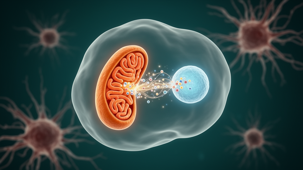
At the heart of the review is a structural claim that has been established in general cell biology: mitochondria and lysosomes communicate through dynamic membrane contact sites, where the two organelles lie closely apposed without fusing. High-resolution microscopy has shown that these sites are dynamic and maintain an average intermembrane distance of approximately 10 nanometres. The small GTPase Rab7 dynamically regulates the formation and dissociation of these contacts, and proteins associated with Rab7, ORP1L and related tethering machinery have been implicated in contact dynamics in non-septic cellular systems. Quantitative super-resolution microscopy has revealed that the endoplasmic reticulum interacts with lysosomal ORP1L via VAMP-associated proteins at mitochondrial division sites, recruiting lysosomes and regulating mitochondrial fission. Other contributors include PDZD8, implicated in contact-site organization in certain cellular contexts, and MAPL, a mitochondrial outer membrane SUMO ligase that promotes the transport of mitochondrial DNA to lysosomes via mitochondria-derived vesicles and contributes to cell death under stress.
The functional consequences of this proximity are substantial. Lysosomal acidification depends on vacuolar ATPases and is influenced by the cell’s energetic state, while lysosomal degradation can recycle metabolites that may indirectly support mitochondrial metabolism. A major downstream consequence is mitophagy, the selective clearance of damaged mitochondria: damaged organelles are sequestered into autophagosomes and degraded after fusion with lysosomes, through pathways including PINK1–Parkin signalling, with hypoxia also activating mitochondrial receptors such as FUNDC1, BNIP3 and BNIP3L. When this communication is intact, dysfunctional mitochondria are efficiently cleared; when it is disrupted, damaged mitochondria accumulate and sustain reactive oxygen species production and inflammatory signalling. Lysosomal acidity is essential for hydrolytic enzyme activity and efficient degradation during this process. The review is careful to note, however, that in sepsis-associated lung injury this functional decoupling should currently be regarded as a strong working model with varying degrees of support across mechanisms and cell types.
Turning to sepsis itself, the authors report that mitochondrial dysfunction is a well-supported feature of the syndrome, documented across multiple experimental systems as well as in human observational studies. Common findings include altered mitochondrial dynamics, loss of membrane potential, reduced ATP production, increased mitochondrial ROS generation and release of mitochondrial danger-associated signals such as mitochondrial DNA. These abnormalities are biologically plausible drivers of inflammatory amplification and tissue injury, and emerging evidence suggests that intracellular LPS triggers caspase-11/GSDMD-dependent mtDNA release, activates cGAS–STING signalling, suppresses YAP1-mediated cell-cycle progression, and impairs endothelial proliferation and vascular repair. Mitochondrial injury also facilitates apoptotic signalling through cytochrome c release and caspase activation. Yet the review stresses that not all aspects of mitochondrial injury have been equally demonstrated in sepsis-associated lung injury specifically, and the precise temporal sequence linking mitochondrial damage to barrier failure and distinct cell death programs in the septic lung remains incompletely resolved.
Lysosomal dysfunction has increasingly been implicated in sepsis as well, particularly in relation to impaired autophagic flux, altered degradative capacity, disturbed inflammatory signalling and defective pathogen handling. When lysosomes fail, damaged mitochondria and invading pathogens are not effectively degraded, leading to intracellular waste accumulation, lysosomal swelling, leakage of lysosomal contents and loss of lysosomal acidity. Selected preclinical studies suggest that improving lysosome-associated homeostasis may support autophagic function and organ protection in sepsis models. The review also highlights immune-mediated regulation of lysosomal homeostasis: interleukin-4 stabilizes lysosomal homeostasis by upregulating lysosomal-associated membrane protein 2 through STAT3 activation, thereby promoting autophagosome–lysosome fusion and alleviating sepsis-induced impairment of autophagic flux, while interferon regulatory factor 7 upregulates autophagy-related genes involved in autophagosome formation and autolysosome maturation, facilitating bacterial clearance in preclinical work. The authors caution that some of these observations derive from broader studies of inflammation and immune cell biology rather than lung-specific sepsis models.
When both organelles falter, the review argues, a vicious cycle can take hold. Mitochondrial and lysosomal dysfunctions are interdependent and mutually reinforcing during sepsis: when mitochondria become dysfunctional, lysosomal α-mannosidase VI mediates mitochondria–lysosome communication, forming a positive feedback loop that exacerbates mitochondrial injury. Defective mitophagy leads to sustained activation of the NLRP3 inflammasome, and systemic inflammation directly damages mitochondria, inducing an energy crisis and excessive ROS production. In experimental sepsis and endotoxin-based lung injury models, impaired mitophagy and disrupted autophagic flux have been associated with mitochondrial ROS accumulation, mtDNA release and enhanced inflammasome signalling. Disrupted crosstalk may also favour the activation of multiple regulated cell death programs, including apoptosis, pyroptosis, necroptosis and ferroptosis, with TLR4-mediated NF-κB signalling considered a potential central hub; in murine models, LPS-induced TLR4-NF-κB signalling may prime pyroptosis by upregulating NLRP3 and pro-IL-1β expression. The review repeatedly emphasizes, however, that the full sequence linking crosstalk failure to NLRP3 activation and lung inflammation remains incompletely mapped in human disease and is based predominantly on preclinical evidence.
On the therapeutic front, the review is deliberately restrained. Sepsis-associated ARDS remains difficult to treat, and current management is still largely supportive. Mitochondria-directed strategies under investigation include preservation of mitochondrial dynamics, reduction of mitochondrial ROS, stabilization of membrane potential and limitation of mtDNA release, while lysosome-directed approaches aim to restore lysosomal acidification, improve autophagic flux and enhance lysosomal biogenesis. Experimental studies have identified candidate regulators in preclinical systems, including TFEB and TBC1D15, and upregulation of transcription factor EB has been reported to reduce inflammatory cytokine expression, promote mitophagy and attenuate lipopolysaccharide-induced lung inflammation and mitochondrial injury in a preclinical model. The authors insist these mechanistic nodes should not be interpreted as therapeutically mature targets: for most candidates the evidence remains preclinical, context-dependent and frequently derived from models that do not fully recapitulate the biological heterogeneity of human sepsis-associated ARDS. No biomarker centred on mitochondria–lysosome crosstalk is currently validated for routine clinical use in sepsis-associated ARDS, although candidate readouts include circulating or alveolar mtDNA, markers of mitochondrial injury, lysosomal enzyme activity and autophagy-related proteins.
The review’s conclusions are measured rather than triumphant. Mitochondria–lysosome crosstalk is described as a compelling organizing framework for sepsis-induced acute lung injury, one that integrates bioenergetic stress, organelle quality control failure, inflammatory amplification and regulated cell death, but the evidence is strongest for some components in general cell stress and experimental sepsis and remains less complete for lung-specific mechanisms and human translational relevance. The authors identify priorities including cell-type-specific mechanistic studies, longitudinal designs that define temporal sequence, human biospecimen validation, spatial multi-omics approaches in human lung tissue and biomarker-guided patient stratification, noting that alveolar epithelial cells, endothelial cells and macrophages are the most studied pulmonary populations while fibroblasts and other stromal or immune subsets remain comparatively underexplored. It also remains unclear which mitochondria–lysosome interactions are causal drivers of injury and which are secondary to inflammation, hypoxia or metabolic stress. For now, the conceptual model is compelling; converting it into credible biomarkers and realistic therapies will require the validation work the authors have laid out.
Subject of Research: Mitochondria–lysosome crosstalk in sepsis-induced acute lung injury
Article Title: Unraveling the mitochondrial–lysosomal crosstalk: a new frontier in sepsis-induced acute lung injury
Article References: Deng, Y., Tu, G., Ma, J., Zhang, Z., Shen, Y., Liu, Y., Wu, J., Deng, Z., Zhong, S., Zou, K., Zhong, B., Zhu, H., & Li, J. (2026). Unraveling the mitochondrial–lysosomal crosstalk: a new frontier in sepsis-induced acute lung injury. Critical Care: Sepsis and Severe Infection, 1(1), Article 15. https://doi.org/10.1186/s44541-026-00011-9
Image Credits: AI Generated
DOI: 10.1186/s44541-026-00011-9
Keywords: sepsis, acute lung injury, ARDS, mitochondria, lysosomes, mitophagy, mitochondrial dysfunction, inflammation, NLRP3 inflammasome, TFEB, narrative review, organelle crosstalk
News Source: Drew Townsend. (October 28, 2026). Mitochondria and lysosomes talk to each other, and sepsis may break that conversation. Scienmag.