


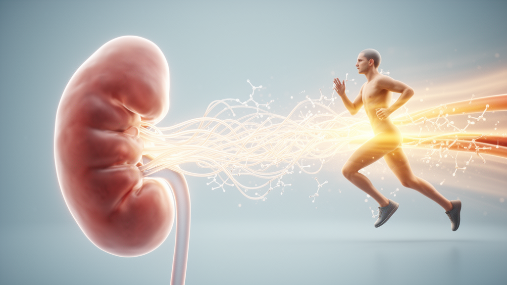
Regular exercise is known to protect the kidney in a range of disease settings, but the molecular machinery behind that protection has remained elusive. A new narrative review published in Frontiers in Physiology argues that microRNAs—tiny RNA molecules that each can regulate dozens of genes at once—may form a crucial regulatory interface between exercise stimuli and renal adaptation. Drawing on studies in rodents and small human cohorts, the authors, led by Binbin Fang of Quanzhou Normal University in China, assemble the scattered evidence into a proposed exercise–miRNA–kidney axis, while candidly acknowledging that direct causal proof is still missing.
The review’s most striking conclusion concerns the strength of the evidence itself. The authors searched PubMed, Web of Science, and Scopus through October 6, 2026, and applied a three-level classification to studies that examined an exercise intervention, one or more microRNAs, and a renal outcome within the same study. Not a single study reached Level A, the tier reserved for work showing that manipulating a specific microRNA alters the kidney’s response to exercise, backed by target validation and rescue experiments. Several studies earned Level B status by linking exercise-induced microRNA changes, a related target pathway, and renal outcomes in the same experiment, while the remainder fell into Level C, representing concurrent associations without pathway validation.
To understand why microRNAs matter for the kidney, the review first maps their pathophysiological roles across disease models. In metabolic and diabetic kidney injury, miR-21 suppresses PPARα-dependent metabolic networks and the mitochondrial protein Mpv17l, impairing fatty acid oxidation and promoting oxidative damage and fibrosis. miR-379 deletion in mice fed a high-fat diet restored the endoplasmic reticulum protein EDEM3 and attenuated albuminuria, glomerular injury, and interstitial fibrosis, while in diabetic kidney disease the same deletion enhanced adaptive mitophagy through FIS1. On the inflammatory front, miR-155 promotes renal inflammation by suppressing SHIP1 and amplifying NF-κB signaling, whereas miR-146a and miR-223-3p act as endogenous brakes, constraining Toll-like receptor and NLRP3 inflammasome pathways.
Cell death and scarring are also under microRNA control. miR-30e-5p suppresses Beclin 1, undermining autophagy and pushing tubular cells toward apoptosis, while miR-182-5p and miR-378a-3p increase ferroptotic susceptibility by targeting the antioxidant defenders GPX4 and SLC7A11. In fibrotic remodeling, the miR-29 family restrains extracellular matrix production, miR-542-3p promotes tubulointerstitial fibrosis through AGO1 repression, and hypoxic tubular cells can package miR-150-5p into extracellular vesicles that activate neighboring fibroblasts via SOCS1 suppression. Perhaps most intriguingly, miR-34a suppresses the longevity-associated protein Klotho, connecting cellular senescence with mitochondrial dysfunction and fibrotic progression in aging kidneys.
Against this backdrop, the review identifies several candidate pathways where exercise, microRNAs, and renal outcomes change together. In aged mice, aerobic exercise reduced renal p53 activation and miR-34a expression, restored Klotho, dampened TGF-β/Smad and Wnt/β-catenin/MMP7 signaling, and reduced renal fibrosis—a coordinated pattern the authors classify as Level B support for a p53–miR-34a–Klotho pathway. In D-galactose-induced renal aging in rats, both moderate-intensity endurance training and high-intensity interval training raised renal miR-296-5p, reduced SGLT2 protein abundance, and attenuated glomerulosclerosis, with high-intensity training also normalizing blood urea nitrogen and serum creatinine.
Diabetic and metabolic models supply further candidates. In diabetic rats subjected to myocardial ischemia–reperfusion injury, exercise alone improved renal histology and catalase activity alongside increased renal miR-126 and Nrf2 expression, earning Level B classification for the miR-126/Nrf2 axis. In mice with high-fat-diet-induced kidney injury, eight weeks of moderate treadmill exercise reduced proteinuria, inflammation, oxidative stress, apoptosis, and collagen deposition, accompanied by broad remodeling of 50 exercise-responsive renal microRNAs; among independently validated candidates, miR-150-5p and miR-182-5p rose with the high-fat diet and fell with exercise. Notably, non-exercise studies show that EV-packaged miR-150-5p drives fibroblast activation and that miR-182-5p promotes ferroptosis, lending biological plausibility to these associations even though those specific pathways were not tested in the exercise study itself.
Exercise dose emerges as a critical variable. In healthy rats, a single bout of exhaustive treadmill exercise increased serum and renal miR-155, reduced SIRT1, activated TXNIP/NLRP3 inflammasome signaling, and produced biochemical and histological signs of acute kidney injury. Chronic moderate training, by contrast, has generally been accompanied by adaptive remodeling—in aged rats, twelve weeks of swimming altered renal miR-21 and miR-34a while restoring PPARα, reducing lipid accumulation, and improving redox balance. The review stresses that exercise intensity, duration, and timing of tissue collection may explain much of the inconsistency across studies, and that renal microRNA responses to exercise do not simply reverse disease-induced dysregulation but may combine normalization, compensation, and exercise-specific adaptation.
One of the review’s most provocative sections concerns skeletal muscle as a potential endocrine-like sender of microRNA signals to the kidney. Extracellular vesicles from damaged or denervated muscle were enriched in miR-21a-3p and aggravated renal injury and fibrosis; in exercise experiments, muscle miR-21a-3p decreased, and plasma vesicles collected after exercise attenuated TGF-β1-induced profibrotic responses in renal tubular cells, though the contribution of that particular microRNA was not directly manipulated. Complementary non-exercise studies show that intramuscular overexpression of miR-23a/27a raised the corresponding microRNAs in serum exosomes and kidney tissue and reduced renal dysfunction and fibrosis in diabetic mice, while muscle-derived exosomal miR-29 and RVG-modified vesicles carrying a miR-23a/27a/26a cluster accumulated in injured kidneys and alleviated tubular injury and fibrosis through validated targets including YY1, TGF-β3, Lpp, Zbtb20, and Klhl42.
Human evidence remains biomarker-focused rather than mechanistic. In patients with nondialysis chronic kidney disease, acute cardiopulmonary exercise raised plasma miR-150 in both patients and healthy participants, while miR-146a fell selectively in the CKD group; after twelve weeks of cycling training, resting concentrations changed little, but the dynamic responses to an acute bout were modified, and a greater exercise-induced reduction in miR-210 tracked with greater gains in peak oxygen uptake. In a cross-sectional study of 63 CKD patients and 18 controls, circulating miR-146a and miR-150 correlated with estimated glomerular filtration rate, arterial stiffness, and exercise capacity, though the capacity associations weakened after adjustment for vascular function. In hemodialysis patients, baseline miR-206 predicted strength gains from intradialytic resistance training, and 24 weeks of training shifted circulating miR-1 and miR-31 alongside improved physical performance and inflammatory measures—associations that, the authors caution, do not by themselves demonstrate improved kidney function.
The overall verdict is one of promising but immature science. Whole-kidney measurements cannot distinguish local transcriptional changes from microRNAs imported from other organs, circulating microRNAs may originate from immune cells, platelets, vessels, or muscle, and no study has yet shown that exercise-responsive vesicle microRNAs actually reach the kidney and mediate protection. The authors call for well-defined exercise interventions combined with microRNA-specific gain- and loss-of-function approaches, direct target validation, rescue experiments, and spatially resolved analyses across glomerular, tubular, endothelial, stromal, and immune compartments. Until such work is done, microRNA remodeling stands as an emerging, plausible, but unproven component of the molecular network through which exercise may safeguard the kidney.
Subject of Research: A narrative review of microRNAs as potential molecular mediators of exercise-induced kidney protection across kidney disease models.
Article Title: Exercise-mediated renoprotection across kidney diseases: emerging roles of microRNAs
Article References: Hong, W., Dong, B., Yu, H., & Fang, B. (2026). Exercise-mediated renoprotection across kidney diseases: emerging roles of microRNAs. Frontiers in Physiology, 17, Article 1981071. https://doi.org/10.3389/fphys.2026.1981071
Image Credits: AI Generated
DOI: 10.3389/fphys.2026.1981071
Keywords: microRNAs, exercise, kidney disease, renal protection, extracellular vesicles, chronic kidney disease, diabetic kidney disease, kidney fibrosis, biomarkers, muscle-kidney communication, Klotho, Nrf2
News Source: Drew Townsend. (October 28, 2026). MicroRNAs May Link Exercise to Kidney Protection, Review Finds. Scienmag.



