A Revolutionary Lens Into the Tumor Microenvironment: Mapping Antibody Distribution at Single-Cell Resolution
The fight against cancer has seen remarkable advances through antibody-based therapies, offering hope especially in hematologic malignancies and certain breast cancers. Yet, this promising avenue stumbles when confronting solid tumors; mere one-fifth of these invasive cancers respond effectively to antibody treatments. A central enigma remains: Do these therapeutic antibodies truly reach their intended cellular targets deep within the dense tumor architecture? Researchers at Stanford Medicine have now unveiled an innovative method that not only reveals the exact whereabouts of antibodies within tumors but also deciphers the intricate cellular interactions influencing their efficacy.
Traditional imaging techniques like positron-emission tomography (PET) scans have offered little more than vague, blurry “hot spots,” indicating the general region of antibody presence but failing to delineate whether the drug is ensconced within the bloodstream, trapped in extracellular matrices, or genuinely engaging cancerous cells. This limitation has hampered the understanding of therapeutic outcomes, leaving clinicians to speculate on the mechanistic reasons behind treatment failure.
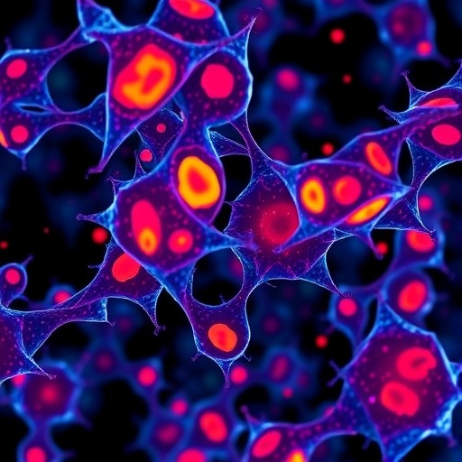
The newly developed approach, termed single-cell spatial pharmacobiology (SSP), merges the power of advanced multiplexed protein detection with precise fluorescent antibody tagging. At its core lies the CODEX technology, pioneered by Professor Garry Nolan’s lab, capable of simultaneously imaging over fifty protein markers within a single tumor tissue slice. When combined with a fluorescent tag attached to the therapeutic antibody, this method maps antibody localization onto a high-definition protein landscape with astonishing precision—aligning cellular boundaries within one micrometer, essentially to the scale of individual subcellular compartments.
Applying SSP to real-world clinical samples from patients undergoing novel fluorescently guided surgeries at Stanford, the researchers uncovered striking heterogeneity in antibody penetration across tumor types. Head and neck squamous cell carcinomas, modest responders to immunotherapy, exhibited relatively higher drug infiltration compared to the notoriously fibrotic and treatment-resistant pancreatic tumors. Even within head and neck tumors, antibody distribution was uneven; only about 16% of cancer cells expressing the epidermal growth factor receptor (EGFR), the antibody’s target, were effectively bound by the drug. These engaged cells predominantly nestled at the tumor periphery adjacent to vascular structures, suggesting a physical bottleneck obstructing drug access to tumor cores.
Delving deeper, analyses revealed that the tumor microenvironment plays a decisive role in modulating antibody delivery. Dense networks of stromal elements — particularly cancer-associated fibroblasts and extracellular matrix proteins like periostin — form formidable physical barricades around tumors. Periostin, a structural protein typically found in tendons and bones, was prevalent in a mesh-like arrangement encapsulating tumor regions with poor antibody penetration. Moreover, a specific fibroblast subtype, capable of producing matrix components including periostin, correlated strongly with these impermeable barriers. This detailed insight uncovers a mechanism by which stromal architecture actively impedes therapeutic efficacy.
Crucially, SSP offers more than just static visualization; it provides a nuanced pharmacological assessment, allowing researchers to determine not only where antibodies accumulate but whether the drug binds to its intended target and exerts anticipated biological effects. Previously, measurement methods relying on plasma antibody levels or radioactive labeling failed to parse such intricate details. By illuminating the precise cellular neighborhoods accessible to drugs, SSP paves the way for rational design of adjunct therapies aimed at dismantling stromal defenses and enhancing antibody delivery.
The implications extend well beyond head and neck or pancreatic cancers. Many solid tumors harbor complex microenvironments whose physical and biochemical landscapes remain poorly understood yet critically influence therapeutic outcomes. With SSP, researchers now have an unprecedented tool to dissect these landscapes comprehensively. It invites a future where cancer treatment becomes highly personalized, informed by spatial pharmacological maps guiding clinicians in predicting response and tailoring combinational strategies.
This breakthrough is also a testament to the transformative potential of interdisciplinary collaboration. The integration of fluorescently labeled therapeutic antibodies from clinical trials with molecular imaging and computational alignment algorithms demonstrates how convergence across clinical oncology, molecular pathology, and bioinformatics can redefine research paradigms. Teams from across renowned institutions including Vanderbilt University Medical Center, Duke University, and international centers contributed their expertise to this pioneering work.
Funding support from the National Institutes of Health and other prominent organizations underscores the importance and promise of this work, propelling forward the frontiers of cancer pharmacology. As SSP technology matures, it holds promise not just for mapping antibody drugs, but potentially other classes of targeted therapies, opening vistas for improved diagnostics, monitoring, and treatment optimization.
By revealing the hidden interplay between therapeutic antibodies and the tumor microenvironment at microscopic resolution, Stanford researchers illuminate a critical bottleneck in cancer treatment. Their innovative single-cell spatial pharmacobiology approach marks a transformative leap toward more effective, tailored immunotherapies that can overcome the stromal barriers thwarting drug delivery. It is a beacon of precision medicine, bringing us closer to fundamentally understanding and conquering solid tumors that have long eluded therapeutic success.
Subject of Research: Cells
Article Title: Single-cell spatial pharmacobiology identifies conserved stromal barriers to therapeutic antibody delivery in human solid tumors
News Publication Date: July 3, 2026
Web References:
https://doi.org/10.1038/s41587-026-03152-x
References:
Lu, G. et al. Single-cell spatial pharmacobiology identifies conserved stromal barriers to therapeutic antibody delivery in human solid tumors. Nature Biotechnology (2026).
Keywords: Cancer cell phenotypes, antibody therapy, tumor microenvironment, spatial pharmacobiology, immunotherapy resistance, extracellular matrix, stromal barriers, fluorescent imaging, CODEX technology, pancreatic cancer, head and neck squamous carcinoma
Tags: antibody-based cancer therapiescellular interactions in tumor antibody efficacyCODEX technology for cancer researchfluorescent antibody tagging techniqueslimitations of PET scans in cancer imagingmapping antibody distribution in tumorsmultiplexed protein detection in oncologyovercoming solid tumor treatment resistanceprecise tumor imaging methodssingle-cell spatial pharmacobiology in cancer treatmentStanford Medicine cancer research innovationstumor microenvironment analysis

