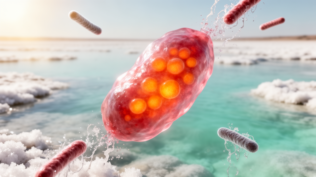
A laboratory study reports the first isolation of the haloarchaeon Halomicrobium mukohataei from Mexico, from brine collected in the Bahía de Lobos coastal lagoon in Sonora. Researchers identified the reddish isolate AS8 by 16S ribosomal RNA gene analysis and tested pigments extracted from its biomass and culture supernatant in antioxidant, antimicrobial and brine-shrimp cytotoxicity assays.
Halomicrobium is a genus of haloarchaea, microorganisms that grow in highly saline environments. The study’s isolate was recovered on medium containing 25% sodium chloride. Haloarchaea can tolerate salt concentrations and other environmental stresses, while producing compounds including carotenoids, exopolysaccharides and antimicrobial substances that have been investigated for biotechnological uses.
The researchers collected 17 brine samples from Bahía de Lobos in April 2022. At the time of collection, the brine measured 27 ± 2 °C, had a pH of 7.6 and had electrical conductivity greater than 20 mS/cm. Cultivation produced 96 haloarchaeal isolates, from which red-pigmented colonies were selected for analysis. The resulting strain AS8 formed circular, slightly mucoid colonies measuring 0.5–2 mm and was described as Gram-negative and able to grow with 25% NaCl.
For molecular identification, the researchers extracted genomic DNA, amplified and sequenced the archaeal 16S rRNA gene, and compared the sequence with records in the NCBI GenBank database. They also performed maximum-likelihood phylogenetic analysis using 14 nucleotide sequences and 1,497 positions. The analysis assigned AS8 to Halomicrobium mukohataei with 100% sequence similarity and an E value of 0.0. The researchers described this as the species’ first report from Mexico and the first study of its pigment activity. They also noted the recognized limitation of 16S rRNA analysis for distinguishing closely related species.
For pigment production, AS8 was grown in liquid medium with 25% NaCl for 10 days with shaking at 150 rpm. The researchers extracted separate biomass pigments and supernatant pigments. The yields were 28 mg per gram of biomass and 140 mg per liter of supernatant, respectively. Thin-layer chromatography showed similar profiles for the two preparations, although the supernatant pigments produced a stronger red-pink signal at an Rf value of 0.45. Fourier-transform infrared spectroscopy showed several shared bands with a β-carotene standard; a band at 1,267 cm−1 attributed to COH vibrations was presented as evidence consistent with bacterioruberin, although the chemical characterization was preliminary.
Both pigment preparations showed concentration-dependent radical-scavenging activity in triplicate DPPH and ABTS assays. In the DPPH assay, the reported scavenging values were 74% for supernatant pigments, 67% for biomass pigments and 38% for the β-carotene control. Corresponding ABTS values were 67%, 52% and 34%. These laboratory assay results indicate greater activity for the tested supernatant preparation than for the biomass preparation and the control under the conditions used; they do not demonstrate effects in people.
In triplicate disk-diffusion tests, both preparations produced inhibition zones against six bacterial species: Bacillus sp., Enterococcus faecalis, Staphylococcus aureus, Escherichia coli, Klebsiella pneumoniae and Pseudomonas aeruginosa. Biomass-pigment inhibition zones ranged from 7 to 13 mm, while supernatant-pigment zones ranged from 7 to 17 mm. Bacillus sp. was the most susceptible organism, with mean zones of 13 ± 1 mm for biomass pigments and 17 ± 0.5 mm for supernatant pigments. Neither preparation inhibited the tested yeast, Candida albicans, under the assay conditions.
The researchers also conducted an animal-based preliminary lethality assay using nauplii of the brine shrimp Artemia salina rather than testing the pigments in humans or cancer cell lines. Biomass pigments produced 40% nauplii mortality at both 0.1 and 1 mg/mL. Supernatant pigments produced 45% mortality at 1 mg/mL and 40% at 0.1 mg/mL. The authors classified both preparations as moderately toxic under the cited classification, while describing the test as an initial screening of possible cytotoxic activity.
Overall, the study provides laboratory evidence that pigments produced by the Mexican AS8 isolate can scavenge assay radicals, inhibit several tested bacteria and cause brine-shrimp mortality. Thin-layer chromatography and infrared spectroscopy suggest carotenoid-like components, including a possible bacterioruberin component, in the extracts. The findings establish biological activity in laboratory assays, but the antimicrobial, antioxidant and cytotoxicity results do not establish clinical efficacy or safety, and the reported work did not test the pigments in people.
Subject of Research: Isolation and laboratory bioactivity testing of pigments from Halomicrobium mukohataei collected in Sonora, Mexico
Article Title: First report of Halomicrobium mukohataei in Mexico and its biological activity
Article References: Cruz-Luna, D., Pina Canseco, S., Hernández Morales, J. L., Aquino Bolaños, T., & García-Sánchez, E. (2025). First report of Halomicrobium mukohataei in Mexico and its biological activity. Neotropical Biology and Conservation, 20(2), 79-92. https://doi.org/10.3897/neotropical.20.e144508
Image Credits: AI Generated
DOI: 10.3897/neotropical.20.e144508
Keywords: Halomicrobium mukohataei, Bahía de Lobos, haloarchaea, microbial pigments, antioxidant assays, antibacterial activity, bacterioruberin, Artemia salina, Mexico, First, report, Halomicrobium
News Source: Drew Townsend. (October 20, 2026). Laboratory study reports first Mexican isolation of Halomicrobium mukohataei and assays its pigments. Scienmag.