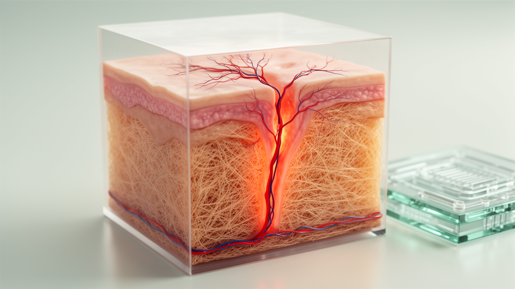
Researchers have built a dynamic, two-cell-type laboratory system that reproduces key features of systemic sclerosis, a connective tissue disease marked by vasculopathy and skin fibrosis. In a preliminary proof-of-concept study published in Arthritis Research & Therapy, endothelial cells grown in a bioreactor and stimulated with immune complexes purified from the sera of patients with systemic sclerosis developed disorganized tubular structures, while fibroblasts cultured downstream in a separate bioreactor showed changes consistent with a fibrotic phenotype.
The system links two bioreactors through a fluidic circuit driven by a peristaltic pump. Endothelial cells were seeded in the apical chamber of a bioreactor containing apical and basal compartments separated by a porous membrane that reproduces the endothelial barrier, with the cells embedded in hydrogel. Skin fibroblasts, isolated from normal healthy subjects, were grown in an acellularized three-dimensional scaffold inside a single-chamber bioreactor. The two devices were then connected so that soluble mediators released by the endothelial cells could reach the fibroblast compartment.
Immune complexes were purified by polyethylene glycol precipitation from the sera of patients with systemic sclerosis who carried anti-centromere or anti-DNA topoisomerase antibodies, as well as from patients with systemic lupus erythematosus and from normal healthy subjects. Endothelial cells were treated with the systemic sclerosis immune complexes, the lupus immune complexes, the healthy-donor immune complexes, the inflammatory cytokine TNFα, or culture medium alone, allowing direct comparison between disease-specific and control stimuli.
When stimulated with the systemic sclerosis immune complexes, endothelial cells in the hydrogel developed unorganized endothelial tubular structures, indicating disruption of normal vascular architecture. The porous membrane proved selective: it prevented the passage of the immune complexes themselves but allowed soluble mediators to cross. This design means any effect seen in the fibroblast compartment could be attributed to soluble signals rather than to direct transfer of the immune complexes.
That downstream effect was evident. Treating the endothelial cells with systemic sclerosis immune complexes induced the formation of fibrous septa in the fibroblast scaffold and increased expression of collagen IV and alpha-smooth muscle actin, while reducing expression of CD34. Together, these changes mirror features associated with fibrotic remodeling of skin tissue, suggesting that signals released from immune-complex-stimulated endothelial cells are sufficient to push fibroblasts toward a profibrotic state.
The soluble mediators measured in the study included the inflammatory and profibrotic signaling molecules IL-6, IL-8, and CCL2, also known as MCP-1. These were quantified, along with the immune complexes, in supernatants collected at different sites along the fluidic circuit, giving the researchers a way to track what was produced where and what crossed the endothelial barrier. Sections of the endothelial cell-hydrogel cultures and the fibroblast-scaffold cultures were evaluated by histological, immunohistochemical, and immunocytochemical analysis.
The authors describe the platform as incorporating five degrees of complexity, hence the label 5D: it is dynamic rather than static, bicompartmental, bicellular, three-dimensional, and includes an endothelial barrier. According to the study conclusions, this innovative dynamic bicompartmental in vitro system allows reproduction of the systemic sclerosis environment, since stimulation of endothelial cells with the disease-derived immune complexes is sufficient not only to disrupt vascular architecture but also to induce skin fibrosis-like changes in the connected fibroblast cultures.
The work was carried out by a team including Elena Raschi, Caterina Bodio, Nicoletta Del Papa, Roberto Caporali, Pier Luigi Meroni, and Cecilia Beatrice Chighizola, among others, and was published open access on 22 October 2026. The article is currently shared as an accepted-manuscript version under a permanent DOI, subject to further editing before the final version of record.
The researchers themselves frame the study as a preliminary proof of concept, and the findings should be read with that scope in mind. The model uses cells and scaffolds rather than living tissue, the fibroblasts derive from healthy donors rather than patients, and the immune complexes come from a defined set of patient sera. Even so, the system offers a controlled way to dissect how immune complexes containing scleroderma-specific autoantibodies, endothelial injury, and fibroblast activation interact, processes that are difficult to separate in patients.
Subject of Research: An in vitro bioreactor model of immune complex-driven vasculopathy and fibrosis in systemic sclerosis
Article Title: A 5D dynamic bicellular in vitro model recapitulates the aetiopathogenic triad of systemic sclerosis: a preliminary proof-of-concept study
Article References: Raschi, E., Bodio, C., Milesi, A., Del Papa, N., Costi, S., Minniti, A., Lonati, P. A., Mauro, A., Borghi, M. O., Caporali, R., Meroni, P. L., & Chighizola, C. B. (2026). A 5D dynamic bicellular in vitro model recapitulates the aetiopathogenic triad of systemic sclerosis: a preliminary proof-of-concept study. Arthritis Research & Therapy. https://doi.org/10.1186/s13075-026-03914-7
Image Credits: AI Generated
DOI: 10.1186/s13075-026-03914-7
Keywords: systemic sclerosis, immune complexes, endothelial cells, fibroblasts, in vitro model, bioreactor, fibrosis, vasculopathy, autoantibodies, IL-6, proof of concept, dynamic
News Source: Ophelia Keating. (October 29, 2026). Lab-Grown Model Reproduces Vascular Damage and Skin Fibrosis of Systemic Sclerosis. Scienmag.