
Indiana University School of Medicine researchers have unveiled a groundbreaking imaging technique that promises to revolutionize the study of bone marrow in preclinical models. This advanced methodological breakthrough overcomes long-standing obstacles in visualizing this complex and crucial tissue, providing unprecedented insight into its cellular architecture while preserving its integrity within the challenging microenvironment of the bone. By enabling detailed, multiplexed visualization of numerous cellular markers simultaneously, this technology sets the stage for transformative advances in understanding diseases rooted in bone marrow dysfunction, including blood cancers, autoimmune conditions, and degenerative musculoskeletal disorders.
Bone marrow, the soft, spongy tissue nestled inside bones, plays a pivotal role in hematopoiesis—the process of blood cell formation—and is crucial for immune system maintenance. Despite its biological importance, detailed investigation of bone marrow microanatomy has been severely limited by its gelatinous nature combined with the rigid encasement provided by the surrounding bone matrix. Traditional imaging modalities have had to contend with either the destructive dissociation of the tissue, as in flow cytometry, or limited multiplex capability in fluorescence microscopy, constraining the scope of molecular and cellular markers that could be concurrently assessed.
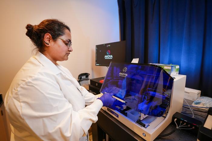
In response to these challenges, the Indiana University team harnessed the power of Phenocycler 2.0™, an advanced multiplex imaging platform that allows for high-dimensional, spatially resolved analysis of tissue specimens. This next-generation instrument was deployed to chart an expansive array of 25 distinct cellular markers within intact mouse bone marrow tissue sections, enabling precise cellular phenotyping without disrupting the native tissue architecture. This level of multiplexing and preservation of tissue context has never before been achieved in bone marrow research, marking a pivotal advance in the field.
The study, which appears in the prestigious journal Leukemia, represents a notable technical leap, as stated by co-lead author Dr. Sonali Karnik. The assistant research professor of orthopedic surgery at IU School of Medicine emphasized that this unique imaging approach not only captures the intricate spatial relationships among diverse bone marrow cell populations but also accesses valuable stem cell niches critical to regenerative medicine and immune function. The technique circumvents the need to mechanically deconstruct tissue for analysis, thereby maintaining native cellular interactions central to understanding disease pathogenesis and therapeutic response.
Prior analytical methods such as flow cytometry, though extremely robust in quantifying cell populations, inherently require cell suspension preparation that destroys the tissue microenvironment and spatial context. Meanwhile, conventional fluorescence imaging techniques typically allow for only a limited number of markers—usually up to three—to be visualized simultaneously. The new multiplex imaging methodology leveraging Phenocycler 2.0 expands this capability nearly ten-fold, offering a comprehensive molecular fingerprint of the bone marrow ecosystem. This technological advantage holds the potential to decode complex pathological mechanisms that underpin hematologic diseases with greater precision.
Importantly, the IU researchers are pioneers in translating the Phenocycler 2.0 platform for mouse bone marrow analysis. While the tool has been previously utilized to image organs such as the spleen and kidney, its application within the dense and delicate bone marrow milieu posed unique challenges. The successful adaptation of this technology opens new avenues for preclinical research, especially in murine models that serve as fundamental platforms for studying human disease mechanisms and therapeutic interventions.
Co-senior author Dr. Reuben Kapur, who directs the Herman B Wells Center for Pediatric Research, highlighted the translational implications of the technique. Mouse models are central to biomedical research due to their genetic tractability and physiological relevance. By enabling detailed, multiplexed imaging of bone marrow in these models, this innovation provides researchers with a potent investigative tool to dissect complex diseases such as leukemia, autoimmune disorders, and other marrow-associated conditions. This capability will likely expedite drug discovery efforts and advance personalized therapeutic approaches.
In anticipation of the broader scientific and commercial applications of this imaging modality, the Indiana University Innovation and Commercialization Office has filed a provisional patent to protect this novel technology. Concurrent with commercialization efforts, research is underway to expand the marker panel to integrate additional components such as bone matrix proteins, neuronal elements, muscular structures, and expanded immune and signaling cell populations. This multifaceted approach seeks to deepen the biological insight obtainable from bone marrow studies, potentially enriching therapeutic target discovery.
The technical sophistication of Phenocycler 2.0 lies in its ability to conduct cyclic immunofluorescence staining and imaging, which involves repetitively labeling tissue with antibodies against different epitopes, imaging, and then chemically or photochemically stripping the labels to allow subsequent rounds. This iterative method enables the detection of an extensive array of biomarkers on the same tissue section with remarkable spatial resolution, preserving cellular and subcellular details. Such multiplex capacity is essential to unravel the heterogeneity and intercellular communications within the bone marrow niche.
Looking forward, the detailed spatial profiling enabled by this technology may offer critical insights into how microenvironmental interactions influence disease initiation, progression, and treatment resistance in hematologic malignancies and immune disorders. Researchers will be better equipped to characterize the dynamic interplay among hematopoietic stem cells, progenitor populations, stromal support cells, and infiltrating immune cells, leveraging the spatial context to inform novel diagnostic and therapeutic strategies.
The collaborative research team contributing to this study includes a multidisciplinary cadre of scientists and clinicians, each bringing specialized expertise in orthopedics, hematology, pathology, and imaging sciences. Their combined efforts underscore the interdisciplinary framework required for such technical innovations to materialize and deliver meaningful biomedical impact. Furthermore, financial support from the National Institutes of Health underpins the project’s significance and potential to drive forward the frontiers of biomedical imaging.
These advancements at Indiana University School of Medicine, the nation’s largest medical school recognized for its extensive NIH funding and innovative research, highlight its leadership in pioneering tools that bridge technological innovation and clinical relevance. Their success in developing a non-destructive, multiplexed bone marrow imaging platform not only opens new research vistas but also sets a precedent for how tissue-based analyses can evolve in the age of high-parameter imaging and precision medicine.
As the scientific community seeks to unravel the complexity of human diseases from their earliest molecular events, methodologies like the one developed at IU represent a vital step forward. This multiplex approach offers unprecedented granularity, spatial context, and biological breadth, allowing researchers to visualize the nuanced cellular environment within bone marrow, ultimately fostering breakthroughs that can translate into improved treatments and patient outcomes.
Subject of Research: Bone marrow imaging and analysis using advanced multiplex imaging technology.
Article Title: Multiplex imaging of murine bone marrow using Phenocycler 2.0™
News Publication Date: 11-Apr-2025
Web References:
Leukemia Journal Article
Indiana University School of Medicine
IU Cooperative Center of Excellence in Hematology
Image Credits: Tim Yates, IU School of Medicine
Keywords: Bone marrow, Blood diseases, Bone diseases, Autoimmune disorders, Cancer treatments
Tags: advanced imaging methodologiesautoimmune condition studiesblood cancer research advancementsbone marrow imaging techniquescellular architecture visualizationdegenerative musculoskeletal disordersdiseases related to bone marrow dysfunctionhematopoiesis and immune systemIndiana University School of Medicine researchinnovative medical imaging technologiesmultiplexed cellular marker analysispreclinical models for bone marrow

