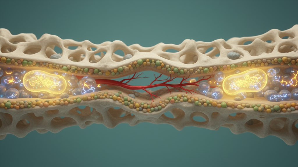
A specialized population of blood vessel cells may hold the key to why some fractures heal poorly, according to a new study published in Cell Death Discovery. Researchers report that an ion channel partnership between the proteins KCNE3 and KCNQ1 coordinates energy production in bone-forming and vessel-forming cells, and that activating this channel with a small molecule called ML277 accelerated fracture healing in mice. The study focused on a vascular subtype marked by high levels of the proteins CD31 and endomucin, known as CD31hiEMCNhi endothelium, which has previously been associated with active support of bone formation.
Using single-cell RNA sequencing, the team identified a subset of these endothelial cells that express the gene Kcne3. These KCNE3-positive cells, found in the epiphysis and periosteum of bone, showed strong stemness markers and appeared to drive differentiation into arterial or sinusoidal endothelium. To probe the channel's function, the researchers silenced Kcne3 in bone marrow stromal cells and in human microvascular endothelial cells grown in the laboratory.
The silencing impaired osteogenic differentiation, angiogenesis, and cell migration. Alongside these functional deficits, the cells showed reduced mitochondrial membrane potential and diminished ATP production, pointing to a breakdown in cellular energy metabolism. The team then generated mice in which Kcne3 could be conditionally knocked out.
These animals lost trabecular bone, had a reduced CD31hiEMCNhi vascular network, and displayed defective fracture healing compared with normal mice. The results link the channel axis to both bone maintenance and the vascular changes that accompany repair. Mechanistically, the study found that fracture healing normally increases potassium ion currents and mitochondrial activity, and that both increases were suppressed when Kcne3 was knocked down.
KCNE3 is known to bind and modulate KCNQ1, a potassium channel, suggesting the pair acts together to couple electrical signaling to mitochondrial energetics during repair. Rescue experiments provided the most striking result. When the researchers activated KCNQ1 channels with the drug ML277 in the Kcne3 knockout mice, osteogenesis, angiogenesis, and bone mass were restored.
This indicated that the downstream channel, KCNQ1, could compensate for the loss of its regulatory partner when pharmacologically stimulated. Beyond the genetic model, ML277 administration in mouse fracture models enhanced trabecular bone volume and promoted the regeneration of CD31hiEMCNhi endothelium, accelerating healing. The findings establish the KCNE3-KCNQ1 axis as a regulator that ties skeletal vascular function to mitochondrial energy production during bone repair.
Because the work relies on mouse models and in vitro cell experiments, its translation to human patients remains to be demonstrated, and the safety or efficacy of channel-targeting drugs for fracture treatment has not yet been tested clinically.
Subject of Research: KCNE3-KCNQ1 ion channel axis orchestrates mitochondrial metabolic reprogramming to couple osteogenesis and angiogenesis in skeletal repair
Article Title: KCNE3-KCNQ1 ion channel axis orchestrates mitochondrial metabolic reprogramming to couple osteogenesis and angiogenesis in skeletal repair
Article References: Wang, R., Wu, Y., Li, Z., Zhao, Y., Gao, Q., Zhou, P., Ji, F., & Hao, T. (2026). KCNE3-KCNQ1 ion channel axis orchestrates mitochondrial metabolic reprogramming to couple osteogenesis and angiogenesis in skeletal repair. Cell Death Discovery. https://doi.org/10.1038/s41420-026-03308-3
Image Credits: AI Generated
DOI: 10.1038/s41420-026-03308-3
Keywords: KCNE3-KCNQ1, channel, axis, orchestrates, mitochondrial, metabolic, reprogramming, couple, osteogenesis, angiogenesis, skeletal, repair
News Source: Daisy Hatcher. (October 24, 2026). Ion Channel Axis KCNE3-KCNQ1 Drives Bone Repair Through Vessel and Energy Metabolism Coupling. Scienmag.