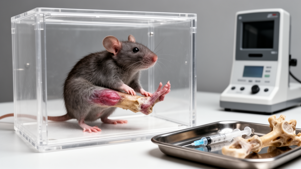
A rat study comparing two experimental approaches to temporomandibular joint osteoarthritis found that intra-articular injection of monosodium iodoacetate, or MIA, produced faster and more severe degeneration of the joint’s condylar cartilage than a model based on unilateral anterior crossbite. Under the tested conditions, the crossbite model generated milder changes that developed progressively, suggesting that the two approaches may be useful for studying different stages and aspects of the disease.
The researchers used 24 healthy adult male specific-pathogen-free Sprague–Dawley rats aged 6–8 weeks and weighing 180–200 grams. After seven days of acclimatization, the animals were randomly and equally divided into three groups: a normal control group, a unilateral anterior crossbite group and an MIA injection group. The study then combined quantitative measurements of condylar height at the anterior, central and posterior regions with histopathological examinations of the temporomandibular joint.
Both experimental models showed signs consistent with osteoarthritis. Safranin O-Fast Green staining, hematoxylin and eosin staining and immunohistochemistry revealed cartilage degeneration, changes in the subchondral bone, fewer chondrocytes and shrinkage of the remaining cartilage cells. Compared with controls, animals in both disease-model groups had significantly higher Mankin and OARSI scores and more positive inflammatory markers. These measurements indicated tissue damage and inflammation, although the investigators did not report the numerical effect sizes or statistical uncertainty for individual outcomes in the supplied abstract.
The principal difference was the intensity and timing of degeneration. The MIA group showed more pronounced increases in Mankin scores, OARSI scores and inflammatory-marker counts than the crossbite group. The authors therefore characterized the injection model as producing more rapid and severe condylar degeneration during the observation period, while the unilateral anterior crossbite model produced a milder, progressively developing form of joint change. The findings provide a direct experimental comparison of the two approaches rather than evidence that either reproduces osteoarthritis in people.
Both models were described as having model-, dose- and time-dependent characteristics, and neither fully captured the complex pathogenesis of human temporomandibular joint osteoarthritis. The results also apply specifically to the male rat population, experimental procedures and observation period used in the study. The authors concluded that model selection should depend on the research question and the disease stage under investigation, rather than on a general ranking of superiority. The study was published as open-access, peer-reviewed research in Scientific Reports, with the supplied version identified as an early publication subject to further editorial changes before the final version of record.
Subject of Research: Experimental comparison of two rat models of temporomandibular joint osteoarthritis
Article Title: Comparative evaluation of unilateral anterior crossbite and monoiodoacetate injection models for temporomandibular joint osteoarthritis in rats
Article References: He, Z., Xiao, Y., Xie, Q., & Jiang, X. (2026). Comparative evaluation of unilateral anterior crossbite and monoiodoacetate injection models for temporomandibular joint osteoarthritis in rats. Scientific Reports. https://doi.org/10.1038/s41598-026-72924-x
Image Credits: AI Generated
DOI: 10.1038/s41598-026-72924-x
Keywords: temporomandibular joint osteoarthritis, rat models, monosodium iodoacetate, anterior crossbite, cartilage degeneration, condylar damage, joint inflammation, histopathology, Sprague–Dawley rats, Comparative, evaluation, unilateral
News Source: Denise Maddox. (October 26, 2026). Injection model caused faster, more severe joint degeneration in rats. Scienmag.