In a finding that could reshape how scientists understand relapses of a common kidney disease, researchers in China have shown that arachidonic acid, a fatty acid released in bulk during infections and inflammatory flare-ups, can dramatically worsen injury to podocytes, the specialized cells that act as the kidney’s final blood-filtration checkpoint. The study, published in the Journal of Cellular and Molecular Medicine, suggests that the damage is driven not just by inflammatory signalling but by a collapse in the cells’ glycolytic energy machinery centred on the enzyme PKM2. When the team pharmacologically reactivated PKM2 in injured podocytes, key metabolic and mitochondrial functions partially recovered, pointing to a potential new therapeutic angle for podocyte diseases.
Podocytes are highly specialized epithelial cells wrapped around the glomerular capillaries, and their interlocking foot processes form the slit diaphragm, a structure essential to the glomerular filtration barrier. When podocytes are injured, proteins leak into the urine, producing the massive proteinuria that defines podocytopathies such as minimal change disease, focal segmental glomerulosclerosis and membranous nephropathy. Clinically, infection is the most common trigger of recurrent proteinuria in these conditions: adults with early-onset, frequently relapsing minimal change disease often develop oedema and heavy protein loss shortly after respiratory infections. Yet even when immunosuppressive and supportive therapies work initially, relapses remain frequent, and the mechanisms by which infection-associated inflammatory signals aggravate podocyte injury have remained incompletely defined.
Arachidonic acid offered a plausible suspect. This omega-6 polyunsaturated fatty acid is stored esterified in cell-membrane phospholipids, and circulating free levels are normally very low, roughly 0.1 micromoles per litre, because the molecule is rapidly bound by albumin. During infection or tissue injury, inflammatory cytokines activate membrane phospholipases that liberate free arachidonic acid, raising its circulating concentration. In kidney disease, proteinuria and hypoalbuminaemia may further reduce albumin’s binding capacity, enlarging the biologically available pool of the fatty acid. Previous work had linked arachidonic acid metabolism to renal inflammation and shown that excessive amounts can harm podocytes, but whether the lipid amplifies damage through metabolic stress in minimal change disease-related injury was unknown.
To find out, the researchers used a puromycin aminonucleoside, or PAN, model, a standard experimental mimic of minimal change disease. Male Sprague-Dawley rats received a single intraperitoneal PAN injection, and ten days later, once podocyte injury was established, half of the injured animals received an intraperitoneal dose of arachidonic acid at 3 milligrams per kilogram. The team was careful to frame this PAN-plus-AA condition as an injury-exacerbation model rather than a faithful reproduction of infection-triggered clinical relapse, since no true infectious stimulus was present. Serum measurements confirmed the design worked: arachidonic acid levels rose from about 4.5 nanograms per millilitre in controls to 8.2 in PAN-treated rats and 12.0 in the combined group, with the combined group also exceeding rats given the fatty acid alone.
The structural consequences were striking. Light microscopy showed no prominent glomerular sclerosis or fibrosis in any group, consistent with the subtle appearance of minimal change disease, so the investigators turned to transmission electron microscopy. There, the glomerular basement membrane thickened from 155 nanometres in controls to 227 nanometres after PAN and 274 nanometres after PAN plus arachidonic acid. Foot-process width swelled from 0.42 to 0.73 and then 1.00 micrometres across the same groups, while the density of foot processes per micrometre of basement membrane fell from 2.04 to 1.16 to 0.81, all differences between the PAN and combined groups statistically significant. Nephrin, the slit-diaphragm protein that anchors the filtration barrier, declined after PAN and dropped further with added arachidonic acid, as shown by both immunofluorescence and Western blotting.
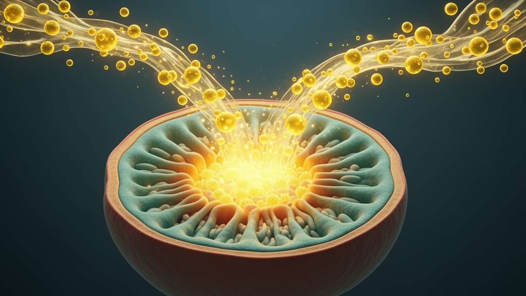
The metabolic story proved equally compelling. Electron microscopy revealed severe mitochondrial injury in podocytes from the combined-treatment rats, including swelling, size heterogeneity, matrix rarefaction and fragmented or dissolved cristae, with quantified increases in mean mitochondrial area, cristae-damage rate and overall damage score. The mitochondrial outer-membrane protein TOM20 was reduced in parallel. The team then examined the PKM enzyme family: PKM1 and PKM2 signals within nephrin-positive glomerular regions both fell after PAN and fell further with arachidonic acid, while phosphorylated PKM2 at the Tyr105 residue rose. Because Tyr105 phosphorylation disrupts formation of PKM2’s active tetramer, the increase signals dysfunction rather than activation. Functionally, total pyruvate kinase activity declined, and renal pyruvate, lactate and ATP levels all dropped, reaching their lowest points in the combined group.
Cultured human podocytes reproduced the pattern. Differentiated cells exposed to PAN alone showed disrupted F-actin stress fibres and reduced nephrin; adding arachidonic acid produced the lowest F-actin intensity and the greatest nephrin loss. The same metabolic signature emerged: reduced PKM1 and PKM2 abundance, elevated phospho-PKM2, diminished pyruvate kinase activity, and lower lactate, pyruvate and ATP. Mitochondria fared worse with the combination, showing greater swelling and cristae damage, a reduced JC-1 aggregate-to-monomer ratio indicating loss of membrane potential, decreased TOM20, and elevated mitochondrial reactive oxygen species detected by MitoSOX staining. Downstream, TUNEL assays revealed more apoptotic cells, and DCFH-DA staining plus flow cytometry confirmed heightened intracellular oxidative stress in the combined-treatment group.
The rescue experiment provided the study’s most translational moment. TEPP-46, a selective PKM2 activator that stabilizes the enzyme’s active tetrameric state, was applied to podocytes under PAN-plus-arachidonic-acid conditions. Treatment restored a more organized actin cytoskeleton, reduced apoptosis, increased PKM2 expression while lowering phospho-PKM2, and raised pyruvate kinase activity along with lactate, pyruvate and ATP production. Mitochondrial readouts improved as well: TOM20 abundance rose, electron microscopy showed less swelling and cristae damage, MitoTracker and JC-1 signals indicated better mitochondrial mass and membrane potential, and ROS levels fell. Together these results indicate that PKM2 activation can partially reverse the metabolic and mitochondrial collapse that arachidonic acid imposes on injured podocytes.
The authors frame their findings within a two-axis model of arachidonic acid toxicity. Their earlier work showed that the fatty acid activates a PKA-c-Abl-nephrin pathway driving actin cytoskeletal remodelling; the present study adds a PKM2-associated metabolic axis that impairs energy production and mitochondrial homeostasis. The two pathways may converge, since actin remodelling demands ATP and mitochondrial ROS can destabilize nephrin and the cytoskeleton, though the data do not establish a strict sequence. Limitations are acknowledged: the model lacks a true infectious trigger, lipidomic analysis did not distinguish free from esterified arachidonic acid or eicosanoid products such as those from COX2/PGE2 signalling, glycolytic flux and respiration were not directly measured by ECAR or OCR assays, and the TEPP-46 rescue was performed only in vitro. Notably, omega-3 fatty acids have recently been reported to protect the podocyte and kidney, underscoring the divergent effects of different polyunsaturated fat subclasses. Even so, the study supports further testing of PKM2-targeted metabolic intervention, including in vivo rescue studies and models that better recapitulate infection-associated relapse, offering hope that a metabolic lever might one day help prevent the proteinuric crashes that follow infections in vulnerable patients.
Subject of Research: Arachidonic acid exacerbation of puromycin aminonucleoside-induced podocyte injury through PKM2-linked glycolytic and mitochondrial dysfunction
Article Title: Arachidonic Acid Disrupts PKM2‐Linked Glycolysis‐Related Metabolism and Exacerbates PAN‐Induced Podocyte Injury
Article References: Nie, K., Gao, X., Liu, M., You, D., Weng, M., Zheng, T., Lin, J., Rao, S., Zhuo, Y., Wan, J., & Yang, L. (2026). Arachidonic Acid Disrupts PKM2 ‐Linked Glycolysis‐Related Metabolism and Exacerbates PAN ‐Induced Podocyte Injury. Journal of Cellular and Molecular Medicine, 30(19), Article e71404. https://doi.org/10.1111/jcmm.71404
Image Credits: AI Generated
DOI: 10.1111/jcmm.71404
Keywords: arachidonic acid, podocytes, PKM2, glycolysis, minimal change disease, mitochondrial dysfunction, puromycin aminonucleoside, nephrin, oxidative stress, TEPP-46, kidney disease, proteinuria
News Source: Jerry Hayes. (October 17, 2026). Inflammatory Fatty Acid Drains Kidney Cells’ Energy Supply, Worsening Podocyte Damage. Scienmag.