


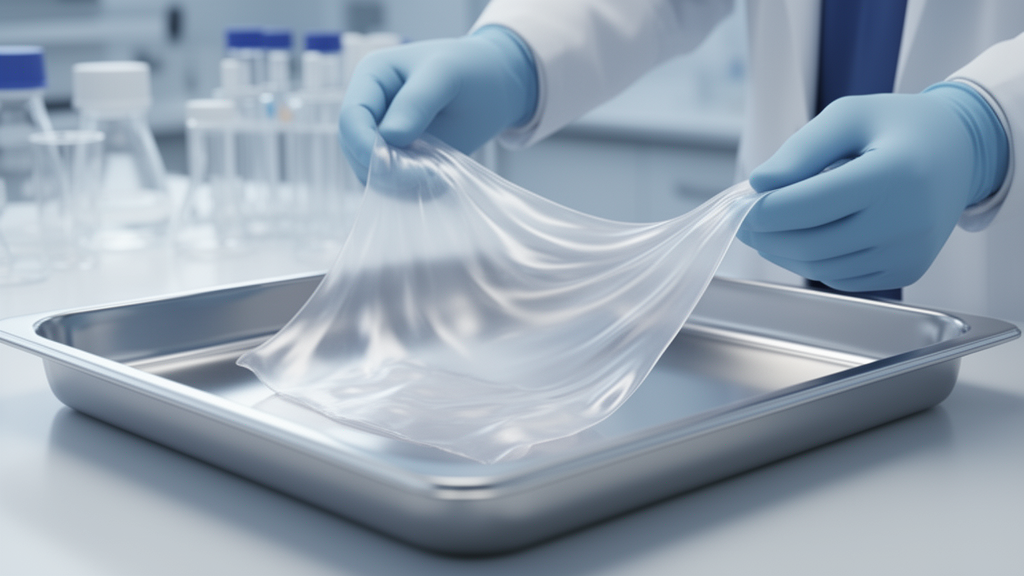
The human amniotic membrane, the innermost natural barrier of the placenta, is moving steadily beyond its long-established role in ophthalmology into orthopaedics, obstetrics and gynaecology, and dermatology. A review published in Cell Regeneration examines how this thin, collagen-rich tissue, typically between 0.02 and 0.5 millimetres thick, is being deployed as a processed biomaterial for tendon repair, osteoarthritis management, prevention of postoperative adhesions, repair of endometrial injury, treatment of diabetic foot ulcers, and burn wound healing. The authors argue that the membrane’s favourable immunological profile and tissue-regenerative properties make it one of the most promising allogeneic repair materials currently available.
Part of the membrane’s appeal lies in what it is made of. Structurally, it consists of an epithelial layer, a basement membrane, and avascular stromal layers, with an extracellular matrix dominated by collagen and harbouring mesenchymal stem cells with multilineage differentiation potential. As a natural allogeneic tissue, it exhibits low immunogenicity, a property attributed largely to immunomodulatory molecules such as human leukocyte antigen-G and factor-related apoptosis ligand. Its epithelial cells and mesenchymal stromal cells secrete interleukin-1 receptor antagonist and tissue inhibitors of metalloproteinases, factors that suppress fibroblast activation and thereby reduce tissue adhesion and scar formation. The same cells release growth factors including basic fibroblast growth factor and hepatocyte growth factor, which promote cell proliferation, migration, and tissue regeneration.
Clinically, the membrane is used in three main processed forms: cryopreserved, dehydrated, and powdered. These formulations have been widely adopted because they are easy to store and handle while retaining key biological functions. The choice of processing matters, however. Comparative studies of anti-adhesion performance after spinal surgery show that freeze-dried membrane resorbs completely within six weeks and provides only temporary barrier protection, whereas crosslinked membrane degrades more slowly, maintains structural integrity longer, and performs on a par with autologous free fat grafts, with better cost-effectiveness and no donor-site morbidity.
In orthopaedics, the review frames the membrane against the limitations of existing tendon repair materials. Synthetic polymer scaffolds such as polycaprolactone and polylactic acid often suffer from low porosity, which restricts cellular infiltration and compromises biological activity. Natural polymer scaffolds frequently lack sufficient mechanical strength, and decellularized extracellular matrix scaffolds are constrained by source scarcity and batch-to-batch variability. The amniotic membrane, by contrast, is rich in collagen, fibronectin, and a broad range of bioactive growth factors, creating a pro-regenerative microenvironment that supports the adhesion, proliferation, and differentiation of tendon stem and progenitor cells. Its mechanical properties can be further enhanced through chemical crosslinking with agents such as polyethylene glycol dialdehyde.
The membrane’s immunomodulatory repertoire is also relevant to tendon healing. Key mediators, including interleukin-10 and thrombospondin 1, regulate the local repair milieu by promoting macrophage polarization toward a pro-healing phenotype, enhancing angiogenesis, and inhibiting matrix metalloproteinase activity. Adhesion prevention is another documented benefit: one clinical study reported that more than 90 percent of patients treated with micronized dehydrated human amniotic membrane injections experienced significant pain relief and reduced adhesion-related symptoms within three months.
For osteoarthritis, injectable amniotic membrane powder has emerged as an alternative to intra-articular therapies such as corticosteroids, platelet-rich plasma, stem cells, and hyaluronic acid, whose long-term disease-modifying effects the review describes as limited. The membrane’s high content of anti-inflammatory mediators, including transforming growth factor-beta and interleukin-10, suppresses pro-inflammatory cytokines in the osteoarthritic joint, alleviating synovitis and slowing cartilage degradation. Preclinical studies have shown that intra-articular administration significantly reduces interleukin-1 beta and tumour necrosis factor-alpha levels while increasing interleukin-10 and interleukin-1 receptor antagonist expression, effectively reversing the pro-inflammatory joint microenvironment. The membrane also upregulates tissue inhibitor of metalloproteinase expression, inhibiting enzyme-mediated degradation of the cartilage extracellular matrix and supporting chondrocyte survival, proliferation, and matrix synthesis. These osteoarthritis findings remain preclinical, and the review does not claim established clinical efficacy for this indication.
In a canine multi-level laminectomy model, glutaraldehyde-crosslinked membrane applied over the dura mater effectively preserved the epidural space. Twelve weeks after surgery, only sparse, loose connective tissue was observed, in contrast to the dense fibrotic adhesions seen in controls. Histological analysis revealed a 51 percent reduction in fibroblast infiltration, significantly decreased collagen deposition, and a markedly attenuated inflammatory response. On this basis, the authors conclude that crosslinked membrane represents a safe, low-immunogenicity biological barrier for preventing postoperative epidural fibrosis, particularly suited to long-segment spinal procedures or patients with insufficient autologous adipose tissue for fat grafts.
Damage to the endometrial basal layer from intrauterine surgery, infection, or other insults frequently leads to intrauterine adhesions, endometrial thinning, and compromised receptivity, while hormonal therapy and surgical adhesiolysis show limited efficacy and high recurrence rates. Unlike passive supports such as collagen sponges or hyaluronic acid gels, the membrane offers a three-dimensional scaffold for cell adhesion, migration, and proliferation, a physical barrier against adhesion formation, and a reservoir of growth factors including epidermal growth factor, vascular endothelial growth factor, transforming growth factor-beta, and interleukin-1 receptor antagonist. In a rat model of endometrial injury, transplantation of membrane-derived mesenchymal stromal cells significantly increased endometrial thickness, glandular density, and vascularization, accompanied by reduced tumour necrosis factor-alpha and upregulated collagen type I alpha 1 chain expression. The authors stress that definitive clinical validation requires large-scale, prospective, randomized controlled trials.
In dermatology, the review covers two applications: treatment of diabetic foot ulcers and promotion of burn wound healing. For diabetic foot ulcers, randomized controlled trials have shown that dehydrated membrane products, used as an adjunct to standard wound care, significantly improve complete ulcer closure rates and reduce healing time compared with standard care alone. The membrane modulates the inflammatory wound microenvironment, downregulating tumour necrosis factor-alpha and interleukin-1 beta when combined with mesenchymal stem cells, and its extracellular matrix component heavy chain-hyaluronic acid/pentraxin 3 suppresses nociceptive neuronal excitation via a CD44 receptor-dependent pathway, blocking pain signalling without the adverse effects of opioids. Experimental studies further show that stem cell-loaded membrane scaffolds accelerate the healing of chronic diabetic wounds in animal models.
In burn care, the membrane serves as a biological dressing that provides a moist physical barrier while delivering bioactive molecules that promote epithelial cell proliferation and migration. Factors secreted by membrane-derived mesenchymal stem cells, including plasminogen activator inhibitor-1, granulocyte colony-stimulating factor, and tissue inhibitor of metalloproteinase-1, suppress heat-induced apoptosis in skin cells through activation of the PI3K/AKT pro-survival pathway. In paediatric burn patients, dehydrated membrane yielded wound healing outcomes equivalent to split-thickness skin grafting while significantly reducing hypertrophic scarring and contracture. Because the membrane’s intrinsic mechanical strength is limited and it degrades rapidly in vivo, modification strategies such as glutaraldehyde crosslinking, which enhances mechanical stability and degradation resistance, and integration with platelet-rich plasma, which increased local growth factor availability and accelerated healing in an animal model, are broadening its clinical applicability. The review concludes that the membrane has evolved from a passive biological dressing into an active regulator of the tissue repair microenvironment, with future work focused on improving mechanical properties for load-bearing repairs, optimizing preservation methods such as glycerination to replace brittle lyophilized products and cold-chain-dependent cryopreservation, and extending applications toward regeneration of nervous and urogenital tissues.
Subject of Research: Applications of human amniotic membrane in tissue repair and regenerative medicine
Article Title: Advances in the application of human amniotic membrane for tissue repair
Article References: Han, J., Yang, X., Khabbir, A., Hu, C., Gao, Y., Liu, X., Liu, R., Zhang, Y., & Lei, Y. (2026). Advances in the application of human amniotic membrane for tissue repair. Cell Regeneration, 15(1), Article 16. https://doi.org/10.1186/s13619-026-00290-0
Image Credits: AI Generated
DOI: 10.1186/s13619-026-00290-0
Keywords: human amniotic membrane, tissue repair, regenerative medicine, orthopaedics, osteoarthritis, endometrial injury, diabetic foot ulcer, burn wounds, biomaterials, immunomodulation, tendon repair, wound healing
News Source: Gregory Coleman. (October 30, 2026). Human Amniotic Membrane Emerges as a Versatile Biomaterial for Tissue Repair. Scienmag.



