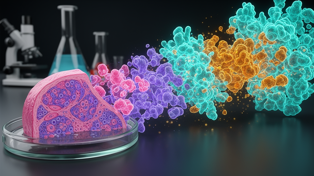
Researchers have developed a deep learning framework called Hist2Prot that infers single-cell spatial proteomic profiles directly from routine hematoxylin and eosin histology images. Spatial proteomics provides high-resolution mapping of protein expression across the tumor microenvironment, but its routine clinical implementation has faced barriers due to cost, technical complexity, and limited tissue coverage. To overcome these constraints, the computational platform reconstructs single-cell protein expression patterns from standard tissue sections commonly used in pathology workflows.
The deep learning model was trained using approximately 51 million paired cells derived from a same-section G4X spatial proteomics dataset. By learning associations between standard cellular morphology and molecular expression, the framework generates virtual single-cell proteomic profiles from standard hematoxylin and eosin slides. The researchers evaluated Hist2Prot across multiple independent testing tiers, including internal test cohorts, independent same-platform G4X cohorts, and external cross-platform datasets totaling approximately 72 million additional cells.
Across these evaluation cohorts, Hist2Prot demonstrated concordance with measured protein profiles and outperformed established baseline methods. The quantitative reconstruction of spatial protein levels operated effectively across diverse cancer types.
The virtual protein measurements generated by Hist2Prot preserved distinct cellular phenotypes within complex tissue architectures. The framework successfully distinguished immune, stromal, and tumor cell subsets based on their reconstructed molecular profiles. Furthermore, the inferred protein distributions recapitulated clinically relevant tumor microenvironment organization.
To assess clinical relevance, the team evaluated the framework across seven independent patient cohorts comprising a total of 1,774 patients. In these validation sets, virtual spatial proteomics enhanced the prediction of molecular biomarkers, patient prognosis, and immunotherapy response compared to models utilizing hematoxylin and eosin morphology alone. Hist2Prot-derived virtual protein profiles and spatial signatures provided predictive utility for therapeutic response beyond standard morphological analysis.
The study authors conclude that Hist2Prot enables scalable, single-cell computational spatial proteomics from routine histology sections for precision oncology.
Subject of Research: Deep learning inference of single-cell spatial proteomics from histology slides
Article Title: The deep learning framework Hist2Prot infers single-cell spatial proteomics from histology to predict biomarkers and clinical outcomes
Article References: Fan, J., Yang, S., Hu, E., An, J., Liu, Q., Liu, N., Ma, M., Prystowsky, M., Lv, T., Yeo, S. Y., Ren, Y., Liu, Y., Li, L., Li, Y., Sun, Q., Qian, P., Lu, L., Deng, W., Pan, X., & Yin, S. (2026). The deep learning framework Hist2Prot infers single-cell spatial proteomics from histology to predict biomarkers and clinical outcomes. Nature Cancer. https://doi.org/10.1038/s43018-026-01270-8
Image Credits: AI Generated
DOI: 10.1038/s43018-026-01270-8
Keywords: Hist2Prot, spatial proteomics, deep learning, computational pathology, hematoxylin and eosin, single-cell analysis, tumor microenvironment, immunotherapy response, precision oncology, biomarker prediction, cancer research, G4X platform, cellular phenotypes, prognostic modeling, tissue morphology, molecular pathology
News Source: Nathaniel Bowman. (November 13, 2026). Hist2Prot Deep Learning Model Predicts Spatial Proteomics Directly From Histology Slides. Scienmag.