Metasurface of self-assembled gold nanoparticles shown to improve resolution of fluorescence images of living cells under a widefield optical microscope to the theoretical limit
Credit: Kaoru Tamada, Kyushu University
In the quest to image exceedingly small structures and phenomenon with higher precision, scientists have been pushing the limits of optical microscope resolution, but these advances often come with increased complication and cost.
Now, researchers in Japan have shown that a glass surface embedded with self-assembled gold nanoparticles can improve resolution with little added cost even using a conventional widefield microscope, facilitating high-resolution fluorescence microscopy capable of high-speed imaging of living cells.
Because optical microscopes magnify light to obtain detailed images of a structure, the size of objects that can be distinguished has long been limited by diffraction–a property of light that causes it to spread when passing through an opening.
Researchers have been developing techniques to overcome these limits with highly advanced optical systems, but many of them depend on the use of strong lasers, which can damage or even kill living cells, and scanning of the sample or processing of multiple images, which inhibits real-time imaging.
“Recent techniques can produce stunning images, but many of them require highly specialized equipment and are incapable of observing the movement of living cells,” says Kaoru Tamada, distinguished professor at Kyushu University’s Institute for Materials Chemistry and Engineering.
Imaging cells using real-time fluorescence microscopy methods, Tamada and her group found that they could improve resolution under a conventional widefield microscope to near the diffraction limit just by changing the surface under the cells.
In fluorescence microscopy, cell structures of interest are tagged with molecules that absorb energy from incoming light and, through the process of fluorescence, re-emit it as light of a different color, which is collected to form the image.
Though cells are usually imaged on plain glass, Tamada’s group coated the glass surface with a self-assembled layer of gold nanoparticles covered with a thin layer of silicon dioxide, creating a so-called metasurface with special optical properties.
Only 12 nm in diameter, the organized metal nanoparticles exhibit a phenomenon known as localized surface plasmon resonance, which allows the metasurface to collect energy from nearby light-emitting molecules for highly efficient re-emission, thereby producing enhanced emission confined to the 10-nm thick nanoparticle surface.
“By introducing the nanoparticles, we have effectively created a light-emitting plane that is only several nanometers thick,” explains Tamada. “Because the light of interest is emitted from such a thin layer, we can better focus on it.”
Additional benefits arise from energy transfer to the metasurface being fast, further localizing emission points by reducing diffusion, and the metasurface’s high refractive index, which helps to improve resolution according to Abbe’s diffraction limit.
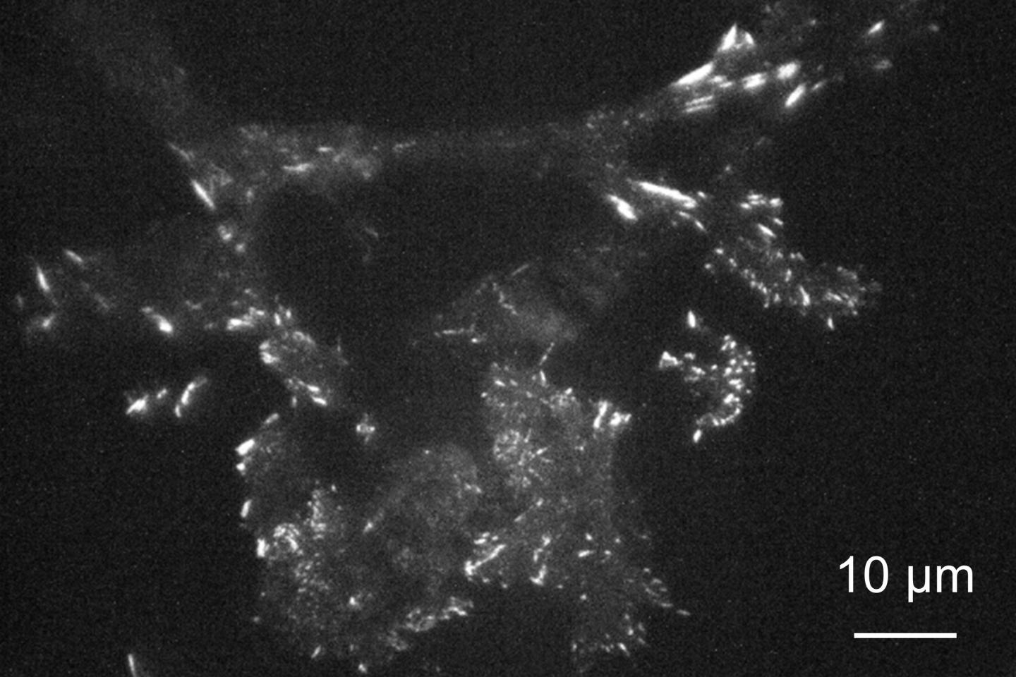
Using the metasurface, the researchers imaged in real-time mouse cells known as 3T3 fibroblasts that were genetically engineered to produce a protein called paxillin that is modified to emit green light when excited. Paxillin plays a key role in creating focal adhesions–points where molecules in the cell membrane interact with the outside world.
Illuminating the entire sample with laser light perpendicular to the surface, the researchers were able to image changes in paxillin near the cell membrane with a higher resolution using the metasurface instead of glass.
Tilting the illumination light to achieve total internal reflection, the researchers could obtain images with even higher contrast because most of the illumination light is reflected off the surface with only a small amount reaching the cell side, thereby reducing stray emission produced by illumination penetrating deep into the cell.
Analysis of images recorded every 500 milliseconds with a super-resolution digital camera revealed clear differences in intensity over spots covering only a few pixels, indicating the resolution was about 200 nm–close to the diffraction limit.
Cells could also be imaged longer on the metasurface because the emission was enhanced despite a lower input energy, thereby reducing cell damage over time.
“Metasurfaces are a promising option for improving resolution for researchers around the world using conventional optical microscopes that they already have,” comments Tamada.
In addition to continuing to improve the surfaces for use with conventional microscopes, the researchers are also exploring the advantages they can have for more sophisticated microscope systems.
###
For more information about this research, see “High axial and lateral resolution on self-assembled gold nanoparticle metasurfaces for live-cell imaging,” Shihomi Masuda, Thasaneeya Kuboki, Satoru Kidoaki, Shi Ting Lee, Sou Ryuzaki, Koichi Okamoto, Yusuke Arima, and Kaoru Tamada, ACS Applied Nano Materials (2020). https:/
This work was supported by JSPS KAKENHI grant number 19H05627 in Japan.
Media Contact
William J. Potscavage Jr.
[email protected]
Original Source
https:/
Related Journal Article
http://dx.


