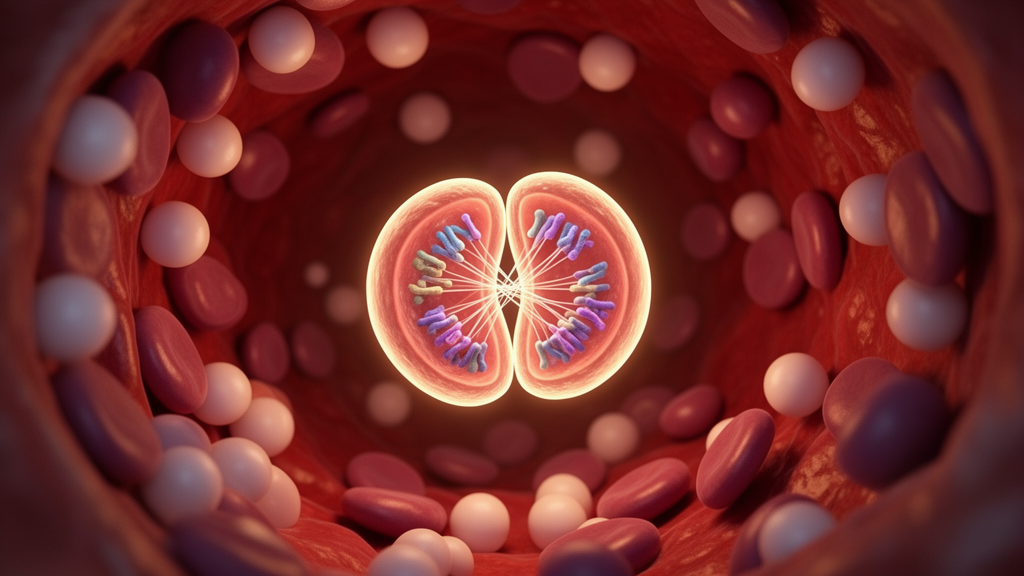
Scientists at the University of Nottingham have identified a previously overlooked mechanism that helps drive acute myeloid leukaemia, one of the most aggressive blood cancers in adults. The discovery centres not on how a mutated protein travels around the cell, the focus of decades of research, but on what happens to that protein during the dramatic process of cell division itself. The findings, published in Nature, reveal that the surface of chromosomes acts as a temporary docking station during mitosis, and that a common leukaemia mutation sabotages this docking system, leaving a key protein stranded in the wrong part of the cell.
Acute myeloid leukaemia, or AML, disrupts the production of healthy blood cells and progresses rapidly, making early and effective treatment critical. Around one-third of patients carry a mutation in a gene called NPM1, one of the most frequently altered genes in this disease. The NPM1 gene provides instructions for a protein that normally resides in the nucleus, the compartment housing the cell’s genetic material. In patients with the mutation, the protein accumulates in the cytoplasm, the surrounding fluid of the cell, and this mislocalisation plays a central role in driving the cancer. Understanding precisely why the protein ends up in the wrong place has therefore been a major goal of leukaemia research.
Until now, most explanations have focused on transport: how the mutated protein shuttles between the nucleus and the cytoplasm. The new study, led by Dr Daniel Booth and Dr Claire Seedhouse at the University of Nottingham’s School of Medicine, shows that this picture is incomplete. Something important happens during cell division itself, when the cell dismantles its nucleus and its chromosomes are exposed in the cytoplasm. The research reveals a previously unknown mechanism that helps explain how the mutation disrupts the normal organisation of proteins inside leukaemia cells.
At the heart of the discovery is the mitotic chromosome periphery, a protective layer that surrounds chromosomes during cell division and has received relatively little scientific attention. Using advanced microscopy, genetic engineering and protein-mapping techniques, the researchers found that healthy NPM1 attaches to this layer during mitosis. This attachment acts like a bookmark: when division is complete and the nucleus re-forms, the protein uses the chromosome surface as a reference point to find its way back to its correct location. In leukaemia cells, this process fails. Dr Booth described the chromosome surface as a temporary docking station that helps proteins return to the right place after a cell divides, and noted that in leukaemia cells this docking does not work properly.
The mutated NPM1 protein behaves in two distinct abnormal ways. It fails to attach properly to the chromosome periphery, instead forming abnormal clusters or dispersing throughout the cell. Worse still, the mutant protein actively interferes with the healthy NPM1 protein produced by the normal copy of the gene, further disrupting its ability to return to the right place after division. Together, these findings reveal two previously unrecognised ways in which cell division contributes to the abnormal positioning of NPM1 in leukaemia, adding a new dimension to a problem that researchers had largely framed as one of transport between cellular compartments.
Perhaps the most exciting aspect of the study is that the researchers showed the process could be partially restored in laboratory experiments. By increasing levels of Ki-67, a protein that helps organise the chromosome periphery, the team created a thicker protective layer around the chromosomes. This improved the attachment of mutant NPM1 to the chromosomes in laboratory-grown leukaemia cells, helping to restore its correct positioning and the normal organisation of the cell. The result suggests that manipulating the chromosome periphery could, in principle, correct one of the fundamental defects that sustains the disease.
The involvement of Ki-67 adds a surprising twist. The protein is widely used in clinics as a marker of cell proliferation and is generally associated with more aggressive cancers, so one might expect higher Ki-67 levels to signal a worse prognosis. However, when the team analysed publicly available patient data, a more complex picture emerged: among patients with this particular form of AML, higher Ki-67 levels showed an unexpected trend towards better survival. The researchers stress that this observation requires further investigation, but it hints that the protein’s role in organising the chromosome periphery may be more protective than its reputation suggests.
Critically, the team confirmed that the newly identified mechanism occurs in samples taken directly from AML patients, not only in laboratory-grown cells. This strengthens the clinical relevance of the findings and suggests that the defective docking of NPM1 at the chromosome periphery is a genuine feature of the disease in patients rather than an artefact of cell culture. Dr Seedhouse said that what is particularly exciting is that the team has shown the process can be improved experimentally, adding that although the work is at an early stage, it reveals a completely new way of thinking about how this form of leukaemia develops and, potentially, how it might be treated.
The discovery challenges the longstanding view that abnormal movement of NPM1 between the nucleus and the cytoplasm is the principal explanation for its mislocalisation. Instead, it identifies interactions between proteins and chromosomes during cell division as another important contributor to the disease. If the findings can be translated into therapies, they would represent a fundamentally new strategy: rather than blocking the mutant protein’s transport, future treatments might aim to strengthen the chromosome periphery so that proteins can find their way home after each cell division. Such an approach could potentially complement existing therapies for AML, a disease in which new treatment options remain urgently needed.
The implications may also extend well beyond leukaemia. The abnormal positioning of proteins inside cells is involved in a range of diseases, and understanding how cells organise their proteins during division could open new avenues of research into other conditions. By uncovering an unexpected link between chromosome biology and blood cancer, the Nottingham team has revealed a new direction for understanding, and potentially treating, diseases driven by misplaced proteins, and has highlighted the mitotic chromosome periphery, long a neglected corner of cell biology, as a promising target for future medicine.
Subject of Research: How the NPM1 mutation disrupts protein positioning at the mitotic chromosome periphery in acute myeloid leukaemia
Article Title: Scientists uncover overlooked mechanism driving aggressive leukemia
Article References: Scientists uncover overlooked mechanism driving aggressive leukemia. (n.d.). Original publication
Image Credits: AI Generated
DOI: Not provided
Keywords: acute myeloid leukaemia, NPM1, cell division, chromosome periphery, Ki-67, protein mislocalisation, University of Nottingham, blood cancer, mitosis, leukaemia research, Nature, mutation
News Source: Nathaniel Bowman. (October 17, 2026). Hidden Cell Division Flaw Emerges as Driver of Aggressive Leukaemia. Scienmag.