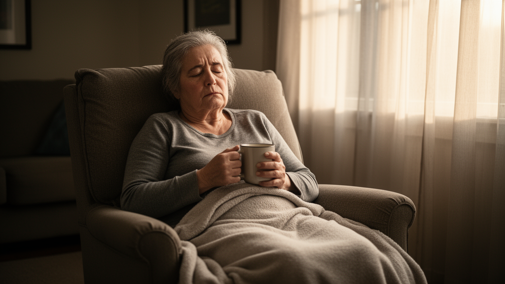
A cross-sectional study of 158 long-term head and neck cancer survivors found that 51.3% met the threshold for clinically relevant fatigue, with a mean score of 44.7 on the European Organisation for Research and Treatment of Cancer Quality of Life Questionnaire Core 30. The average exceeded the age- and sex-adjusted score of 29.7 reported for the German general population. Fatigue was closely associated with poorer health-related quality of life, greater psychological distress and stronger fear of cancer recurrence, while radiation exposure to selected cerebral regions showed no association that remained significant after correction for multiple testing.
The participants had received curative radiotherapy at University Medical Center Leipzig, Germany, between 2005 and 2023 and were surveyed between January 2024 and January 2025. Their median age at the beginning of radiotherapy was 60 years, and the median interval between completing treatment and entering the study was 41 months. Most cancers were in the oropharynx, which accounted for 38.6% of the cohort, followed by the oral cavity at 28.5% and the larynx at 13.9%. All participants had received intensity-modulated radiotherapy or volumetric modulated arc therapy. Of the 158 people included in the analysis, 88, or 55.7%, also received concurrent systemic treatment; 41 underwent definitive chemoradiotherapy and 47 postoperative chemoradiotherapy.
Fatigue was associated with functional and psychological measures, although the observational design cannot determine whether these relationships were causal. The strongest correlation with quality of life was an inverse association with physical functioning, for which the Spearman coefficient was −0.72. Fatigue also correlated inversely with role functioning at ρ = −0.66 and with cognitive functioning at ρ = −0.46. Overall health-related quality of life was inversely associated with fatigue at ρ = −0.59, while psychological distress was positively associated at ρ = 0.54 and fear of cancer recurrence at ρ = 0.41. All three relationships had statistical significance values below 0.001. The head and neck-specific quality-of-life scales were also significantly correlated with fatigue, with correlations ranging from −0.72 for physical functioning to −0.46 for cognitive functioning and from 0.20 for diarrhea to 0.60 for pain and dyspnea among symptom scales.
Clinical factors were also examined. Survivors whose Eastern Cooperative Oncology Group performance status was impaired at the start of radiotherapy reported more fatigue than those with a performance status of zero. Their mean scores were 49.9 and 40.2, respectively, in an analysis with p = 0.03. Fatigue also varied by tumor site, with particularly high scores among patients with hypopharyngeal cancer and laryngeal cancer. The mean score for the hypopharyngeal group was 74.6, compared with 51.5 for the laryngeal group. No significant associations were found for age, comorbidity burden as measured by the Charlson Comorbidity Index, time since radiotherapy, smoking status, tumor stage, p16 status, type of radiotherapy, concurrent systemic treatment, neck irradiation or recurrence. The site differences may reflect underlying variations in performance status, smoking or comorbidity rather than an effect of tumor location itself.
The investigation also tested whether radiation reaching cerebral organs at risk might be associated with persistent fatigue. Researchers retrospectively delineated the brainstem, cerebellum, posterior fossa, medulla, pituitary gland, basal ganglia and hippocampus, then recalculated treatment plans and examined both conventional dose metrics and equivalent doses in 2-Gy fractions. The brainstem, for example, had a median mean equivalent dose of 3.59 Gy, while the corresponding values were 5.65 Gy for the cerebellum, 5.36 Gy for the posterior fossa and 11.53 Gy for the medulla. Maximum doses were higher in some structures, but five of 18 univariable correlations reached only nominal statistical significance, and none survived the study’s Holm-Bonferroni correction across all 18 tests. Mean and maximum doses showed a similar pattern, and the volume receiving at least 95% of the prescription dose was not correlated with fatigue, at r = 0.03 and p = 0.69.
The researchers also conducted voxel-based analysis, registering CT and dose data to a common reference image to test dose-outcome relationships one voxel at a time. Small to moderate clusters in the right cerebellum initially suggested a possible relationship between local dose and fatigue, but no voxel remained significant after permutation-based correction for the family-wise error rate. The analysis therefore did not identify a cerebral dose pattern robustly associated with fatigue in this cohort. With 154 participants available for the reported dosimetric precision calculation, the study could detect a correlation of |r| ≥ 0.22 with 80% power at a two-sided significance level of 0.05, meaning weaker associations could not be excluded.
Baseline skeletal muscle mass was also not associated with fatigue. A deep-learning segmentation model was used to quantify skeletal muscle area on CT scans taken at the middle of the third cervical vertebra, and this measure was converted into a skeletal muscle mass index. Its correlation with fatigue was weak and nonsignificant, at r = −0.05 with a 95% confidence interval from −0.21 to 0.11. Because muscle mass was measured at the beginning of radiotherapy and fatigue was assessed a median of 41 months later, the baseline value may not have represented the survivors’ muscle status at the time of the survey. The result does not rule out sarcopenia as a contributor, particularly if muscle health changed during or after treatment.
Several possible explanations were offered for the lack of a radiation-dose relationship. The three-item fatigue scale used in the core quality-of-life questionnaire primarily captures physical fatigue and less fully represents emotional and cognitive fatigue, potentially missing associations that a dedicated fatigue instrument might detect. The study population was also clinically heterogeneous, whereas earlier studies reporting links between brain dose and fatigue often focused on more uniform groups, including patients with nasopharyngeal cancer. In addition, the relatively low median cerebral doses in this cohort may have limited the range in which a dose effect could be detected. Because every participant received IMRT or VMAT, the study could not compare these techniques with older three-dimensional conformal radiotherapy. A prior voxel-based analysis also reported a right-posterior cerebellar association in one of two participating centers; the present study found a similar region, but its result did not remain significant after correction for multiple testing.
Several limitations constrain the findings. The study was single-center and cross-sectional, so it did not track changes in fatigue from before treatment through survivorship or establish whether fatigue preceded changes in distress and fear of recurrence. No a priori sample-size calculation was performed, although the authors’ precision analysis indicated that weaker correlations could not be excluded. Psychosocial interventions and supportive care were not documented, blood tests were not performed, and possible contributors such as anemia and hypothyroidism were not assessed. The study used the three-item fatigue scale rather than a dedicated questionnaire covering physical, emotional and cognitive fatigue in greater detail. Most analyses other than the prespecified dosimetric and voxel-based corrections were exploratory, and their p-values were reported descriptively without multiple-testing correction.
Participation may also have affected the composition of the cohort. Of 303 patients approached, 202 returned completed questionnaires, but 44 participants were subsequently excluded, leaving 158 for analysis. Compared with participants, non-responders more often had an ECOG performance status of at least 1, less often received concurrent systemic treatment and more often experienced recurrence after radiotherapy. These differences suggest that the included group may not represent all survivors, particularly those with poorer functional status or a less favorable clinical course. Overall, the results identify fatigue as a substantial burden in the studied head and neck cancer population, affecting about half of participants and closely associated with impaired physical functioning, lower quality of life, distress and fear of recurrence. No tested cerebral dose metric was robustly associated with fatigue, and baseline muscle mass was not associated with it either.
Subject of Research: Prevalence, associated factors and cerebral radiation-dose correlates of fatigue in head and neck cancer survivors after radiotherapy
Article Title: Fatigue in head and neck cancer survivors after radiotherapy – prevalence, risk factors, and radiation dose correlates
Article References: Magnus, V., Schäfer, S., Tyyger, M., Iyizoba-Ebozue, Z., Unger, D., Tews, A.-M., Boide, A., Mehnert-Theuerkauf, A., Seidel, C., Moustakis, C., Nicolay, N. H., & Rühle, A. (2026). Fatigue in head and neck cancer survivors after radiotherapy – prevalence, risk factors, and radiation dose correlates. Journal of Cancer Survivorship. https://doi.org/10.1007/s11764-026-02143-w
Image Credits: AI Generated
DOI: 10.1007/s11764-026-02143-w
Keywords: head and neck cancer, cancer survivorship, radiotherapy, chronic fatigue, quality of life, psychological distress, fear of recurrence, radiation dosimetry, skeletal muscle, cross-sectional study, Fatigue, head
News Source: Nathaniel Bowman. (October 20, 2026). Half of head and neck cancer survivors report clinically relevant fatigue. Scienmag.