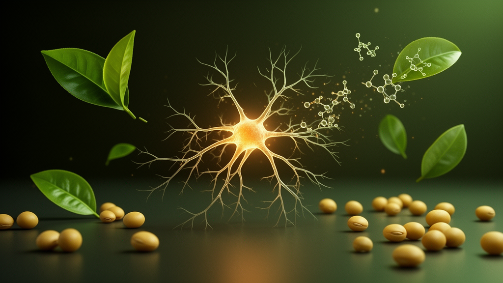
Two plant-derived compounds, one from green tea and one from soybeans, have shown a broad set of protective effects in laboratory models relevant to Alzheimer’s disease, according to a new study published in Frontiers in Natural Products. Researchers at Anurag University in Hyderabad, India, compared epicatechin gallate (ECG), a flavan-3-ol catechin abundant in green tea, with genistin (GIN), the predominant glycosylated isoflavone in soybeans, across a battery of biochemical assays and two cell-based models. Across nearly every test, ECG outperformed GIN, although both compounds displayed measurable activity against several pathological processes linked to the disease.
The study was motivated by the multifactorial nature of Alzheimer’s disease, which involves cholinergic dysfunction, oxidative stress, neuroinflammation, and the accumulation of amyloid-beta (Aβ) peptide. Because no single mechanism accounts for the disorder, the authors argued that compounds capable of acting on multiple pathways simultaneously deserve systematic comparison. Previous work had examined GIN and ECG individually in different experimental settings, but the two had not been evaluated side by side within a single framework covering cholinesterase inhibition, antioxidant capacity, Aβ-induced neurotoxicity, and inflammatory nitric oxide production.
The cholinesterase experiments used Ellman’s colorimetric method in 96-well plates, testing concentrations from 1 to 30 micromolar. Both compounds inhibited acetylcholinesterase (AChE) and butyrylcholinesterase (BuChE) in a concentration-dependent manner. ECG was the stronger inhibitor of both enzymes, with an AChE IC50 of 12.85 ± 0.75 micromolar compared with 17.13 ± 0.79 micromolar for GIN, while the reference drug donepezil remained more potent at 8.28 ± 0.34 micromolar. The same ordering held for BuChE, where ECG recorded an IC50 of 14.44 ± 0.67 micromolar against 18.72 ± 0.84 micromolar for GIN, with rivastigmine as the standard. At the highest concentration of 30 micromolar, ECG inhibited AChE by 91.8 percent and BuChE by 91.5 percent.
Antioxidant performance was assessed with five complementary assays: DPPH and ABTS radical scavenging, ferric reducing antioxidant power (FRAP), a superoxide dismutase-like assay, and a catalase activity assay. ECG again showed consistently stronger activity than GIN, with lower IC50 or EC50 values in every test. Notably, in the FRAP assay ECG exceeded even ascorbic acid, reaching an EC50 of 15.81 ± 0.90 micromolar versus 18.54 ± 1.10 micromolar for the vitamin, while GIN trailed at 22.20 ± 1.30 micromolar. The authors attributed ECG’s advantage to its galloyl moiety, which carries three additional phenolic hydroxyl groups and a carboxyl group that may enhance interactions with reactive species, although they cautioned that the structural basis of the difference requires further validation.
To model neuroprotection, the team used undifferentiated SH-SY5Y human neuroblastoma cells injured with synthetic Aβ1–42 oligomers at a final concentration of 10 micromolar for 24 hours. Microscopy revealed characteristic damage in the Aβ-treated cells, including widened gaps between cells, reduced cell-cell connections, rounding and shrinkage, and lower overall cell density. Co-treatment with either compound partially reversed these changes, with ECG-treated cultures showing better cell adherence, improved connectivity, and more preserved elongated neuronal morphology than GIN-treated cultures at matched concentrations.
Quantitative viability measurements supported the visual findings. Aβ exposure alone reduced SH-SY5Y viability to roughly 42 to 44 percent, whereas co-treatment with ECG restored viability to 97.67 ± 5.1 percent at 10 micromolar and 96.49 ± 4.8 percent at 25 micromolar, compared with 94.28 ± 4.7 percent and 94.01 ± 4.5 percent for GIN at the same doses. Both compounds alone maintained viability of about 92 to 100 percent across a wide 1 to 150 micromolar range, indicating good cytocompatibility. Lipid peroxidation, measured as malondialdehyde (MDA) by the thiobarbituric acid-reactive substances method, rose to 3.95 ± 0.22 nanomoles per milligram of protein in Aβ-injured cells from a baseline of 1.25 ± 0.05, but fell to 1.78 ± 0.07 with ECG co-treatment and 2.05 ± 0.19 with GIN, indicating attenuation of oxidative damage.
Anti-inflammatory activity was tested in BV-2 murine microglial cells stimulated with lipopolysaccharide (LPS) at 1 microgram per milliliter. Before the inflammation assays, both compounds maintained BV-2 viability above 95 percent even at the highest tested concentration of 150 micromolar, with no concentration-dependent cytotoxicity. In the nitric oxide inhibition assay, both GIN and ECG suppressed LPS-induced nitric oxide production in a concentration-dependent manner across 1 to 50 micromolar, although neither matched the reference inhibitor L-NAME, which achieved 83.6 ± 3.9 percent inhibition at 50 micromolar versus 75.8 ± 3.7 percent for ECG and 68.9 ± 3.4 percent for GIN. The authors emphasized that nitric oxide was the only inflammatory marker measured, so the results demonstrate NO-suppressive activity rather than a comprehensive characterization of anti-inflammatory effects.
The study’s authors were careful to spell out the limits of what these findings mean. The in vitro biochemical assays assessed free radical scavenging and reducing capacity under chemical conditions and do not directly reflect intracellular antioxidant activity. The use of undifferentiated SH-SY5Y cells, which do not fully represent mature neurons, was acknowledged as a limitation, as was the qualitative nature of the morphological assessment and the limited specificity of the TBARS method for MDA. Importantly, the researchers stressed that the concentrations effective in their cell models cannot be directly extrapolated to achievable brain concentrations, because oral bioavailability, metabolism, pharmacokinetic exposure, and blood-brain barrier penetration were not evaluated.
Statistically, all experiments were performed in triplicate with three independent biological replicates, analyzed by one-way ANOVA with Tukey’s multiple-comparison testing, and concentration-response curves were fitted using four-parameter logistic nonlinear regression with 95 percent confidence intervals. The authors concluded that GIN and ECG may be considered promising experimental candidates for further investigation, while noting that the precise molecular mechanisms underlying the observed effects were not investigated and that mechanistic studies and appropriate in vivo investigations are required to determine their biological relevance and potential applicability to Alzheimer’s disease.
For readers following the search for multitarget approaches to neurodegeneration, the study offers a structured head-to-head comparison rather than a therapeutic claim. Its value lies in consolidating evidence that two widely consumed dietary phytochemicals can modulate several disease-associated processes in simplified laboratory systems, with ECG showing comparatively stronger activity across most endpoints. Whether these effects translate to the far more complex environment of the human brain remains an open question that only properly designed animal and, eventually, clinical studies can answer.
Subject of Research: Laboratory evaluation of the phytochemicals genistin and epicatechin gallate against cholinesterase activity, oxidative stress, amyloid-beta neurotoxicity, and nitric oxide production in models relevant to Alzheimer's disease.
Article Title: Modulation of cholinergic dysfunction, oxidative stress, and neuroinflammation by selected phytochemicals: neuroprotective evidence from in vitro biochemical and cell-based models of Alzheimer’s disease
Article References: D, R. S., & Pathakala, N. (2026). Modulation of cholinergic dysfunction, oxidative stress, and neuroinflammation by selected phytochemicals: neuroprotective evidence from in vitro biochemical and cell-based models of Alzheimer’s disease. Frontiers in Natural Products, 5, Article 1945279. https://doi.org/10.3389/fntpr.2026.1945279
Image Credits: AI Generated
DOI: 10.3389/fntpr.2026.1945279
Keywords: Alzheimer's disease, epicatechin gallate, genistin, phytochemicals, cholinesterase inhibition, oxidative stress, SH-SY5Y cells, BV-2 microglia, nitric oxide, amyloid-beta, in vitro study, neuroprotection
News Source: Cassandra Pierce. (October 27, 2026). Green Tea and Soy Phytochemicals Show Neuroprotective Effects in Lab Models of Alzheimer’s Disease. Scienmag.