Synaptic dysfunction is identified as a critical early feature in the progression of Alzheimer’s disease and Parkinson’s disease, often preceding the extensive neuronal loss observable through conventional imaging. Impairments in long-term potentiation, long-term depression, and metaplasticity have been shown to emerge from convergent biochemical disruptions at the postsynaptic density. These alterations, which include distorted receptor trafficking, aberrant synaptic protein phosphorylation, and mitochondrial stress, are linked to cognitive and motor decline. Because current pharmacological and neuromodulatory interventions are often limited by their inability to restore the underlying nanoscale biochemical codes that govern plasticity, a new narrative review examines the conceptual possibility of using graphene-based neuromorphic scaffolds functionalized with specific phosphoprotein motifs to bridge these disease-specific signaling gaps.
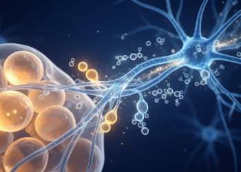
The postsynaptic density is a complex nanodomain where structural proteins, kinases, scaffold complexes, and ligand-gated receptors coordinate to translate synaptic inputs into biochemical programs. In healthy neural circuits, the N-methyl-D-aspartate receptor-CaMKII-PSD-95 signaling triad is essential for maintaining long-term potentiation and memory consolidation. In Alzheimer’s disease, amyloid-beta oligomers aberrantly bind to NMDAR subunits, promoting excessive calcium influx and PSD-95 proteolysis. Concurrently, tau pathology disrupts Fyn-mediated coupling between receptors and scaffolds. In Parkinson’s disease, reduced dopaminergic signaling through D1 receptors results in decreased phosphorylation of glutamate receptor substrates such as GluA1, further dismantling the capacity for activity-dependent synaptic strengthening and leading to network-level shifts toward synaptic weakening.
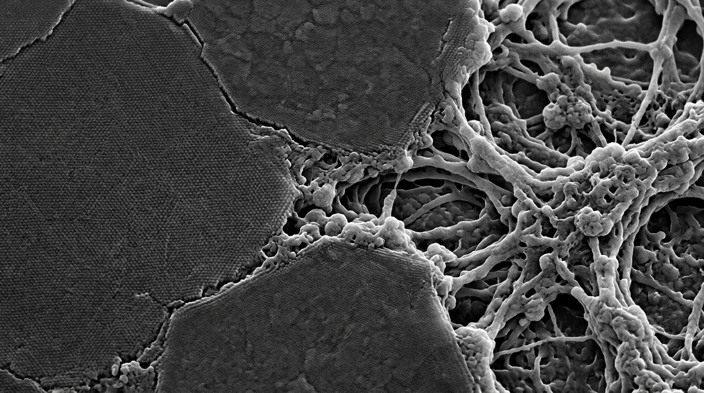
Recent neuroproteomic studies have uncovered quantifiable plasticity signatures involving the CaMKII-NMDAR-PSD-95 axis, AMPAR subunit phosphorylation, and kinase-phosphatase network recalibration. These findings suggest that the synaptic phosphoproteome becomes disordered long before overt synaptic loss occurs, establishing a potential window for intervention. Parallel advances in graphene-based neuromorphic devices have demonstrated programmable conductance states and electrical responses capable of emulating aspects of spike-timing-dependent plasticity. The proposed framework investigated in the review involves functionalizing graphene surfaces with synthetic phosphoprotein motifs to impart molecular selectivity to these artificial interfaces, theoretically allowing them to recognize endogenous receptor complexes or reproduce disease-specific plasticity rules.
Despite the potential for integrating molecular signatures with neuromorphic engineering, the review emphasizes that electronic emulation of plasticity-related signaling has not established the restoration of endogenous phosphorylation networks or therapeutic efficacy in neurodegenerative disease models. The biochemical-to-electronic correspondence remains a design hypothesis rather than a demonstrated functional repair. The proposed device architecture would require an explicit, experimentally calibrated transfer function connecting molecular recognition to an ionic or electrical input, and subsequently to conductance transitions and retention times. Current materials engineering for neural interfaces must distinguish between device-level conductance updates and the actual biological regeneration of synaptic signaling machinery.
Neuromodulator-dependent pathways, such as those involving acetylcholine and dopamine, further complicate the design of a synaptic prosthesis. These inputs modify how neurons interpret glutamate-driven plasticity signals. Acetylcholine-driven M1 muscarinic receptor signaling, involving PKC and the beta-PIX-Rac1-PAK pathway, and glutamate-driven NMDAR-dependent signaling, which involves CaMKII and the ARHGEF2-RhoA-ROCK-SHANK3 pathway, are established examples of these regulatory networks. A graphene transistor does not inherently detect receptor activation or intracellular phosphorylation. Therefore, the interface would require validated receptor- or phosphosite-selective capture elements to function within the broader regulatory network, rather than operating as a self-sufficient molecular code.
The review identifies several translational limitations that must be resolved before disease-specific synaptic repair can be evaluated. These include the necessity for demonstrating selective molecular binding, stability in biological media, and temporal fidelity of the proposed hybrid system. Chronic compatibility is also a significant barrier; graphene derivatives and related materials require rigorous testing to ensure they do not provoke reactive gliosis, biofouling, or fibrotic encapsulation. The long-term performance of these devices, including potential material degradation and electrical drift, remains unknown in the context of neurodegenerative disease environments where cellular homeostasis is already under severe stress.
Establishing a convincing biological correspondence would require concurrent phosphosite measurements, receptor trafficking assays, and electrophysiological recordings in the same experimental model. Without such data, electrical conductance changes in graphene devices cannot be interpreted as a replacement for damaged molecular decision pathways. Furthermore, synapse-directed therapy must account for cell-type-specific neuromodulator input, inhibitory circuitry, and glial interactions, none of which can be reduced to a single conductance threshold. The review concludes that while the convergence of phosphoproteomics and graphene engineering offers a scientifically plausible framework, it remains a testable concept that requires stepwise experimental validation before any clinical application can be considered.
The evidence hierarchy presented by the authors clarifies that the current state of research is exploratory. Initial work must establish reproducible molecular functionalization and phosphosite discrimination, followed by device-level electrochemical characterization and neuronal co-culture experiments. Only after these hurdles are cleared could network or behavioral outcomes, such as spatial learning or motor-sequence performance, be evaluated. The review explicitly cautions that it does not assert the efficacy of an experimentally unvalidated synaptic prosthesis, highlighting instead the priority of mapping region- and cell-type-specific signaling to guide future, more precise bioelectronic designs.
Beyond the primary scaffold-receptor collapse, the cytoskeletal architecture of dendritic spines represents a vital, phosphorylation-dependent structural correlate of synaptic strength that is fundamentally compromised in these neurodegenerative states. The actin-severing protein cofilin, which is tightly regulated by inhibitory phosphorylation at its Ser3 residue, becomes aberrantly dephosphorylated in both Alzheimer’s and Parkinson’s disease. This loss of inhibitory control promotes premature actin depolymerization, leading to the selective atrophy of thin, highly plastic spines that are essential for episodic memory formation and motor skill refinement. Concurrent failures in drebrin and WAVE complex regulation further prevent spine maturation, resulting in a reduced postsynaptic surface area that exacerbates the instability of the entire signaling apparatus. Engineering approaches that do not account for the structural requirement of stable cytoskeletal anchorage may fail to restore functional synapse geometry, as mere receptor recruitment without structural scaffold reinforcement remains insufficient for physiological synaptic persistence.
Metabolic failure and redox imbalances further diminish the fidelity of synaptic transmission, complicating the restoration of plasticity through any bioelectronic interface. Mitochondria, which act as the primary engines for the energetically expensive processes of neurotransmitter release and ion pumping, often show early signs of fragmentation and trafficking defects in affected neurons. Insufficient ATP levels impair the activity of key enzymes and ion transporters, leading to synaptic exhaustion and an inability to stabilize the potentiated states required for long-term memory. These bioenergetic deficits occur in tandem with presynaptic terminal dysfunction, where the phosphorylation states of synapsins and Rab3 GTPases are distorted. In Parkinson’s disease, the pathogenic accumulation of α-synuclein physically disrupts the assembly of SNARE complexes, reducing the probability of synchronized glutamate release. These presynaptic errors break the timing precision required for spike-timing-dependent plasticity, fundamentally decoupling the presynaptic drive from the postsynaptic response.
The complexity of neuromodulatory signaling adds additional design requirements for any proposed synaptic interface. Acetylcholine-dependent M1 muscarinic receptor signaling, which engages the PKC-β-PIX-Rac1-PAK pathway, provides a necessary context for spine remodeling that is distinct from glutamate-driven excitation. Similarly, the NMDAR-dependent CaMKII pathway coordinates with the RhoA-ROCK-SHANK3 axis to translate neuronal activity into structural modification. These specific molecular cascades show that plasticity is not a singular binary switch, but a multi-input integration process influenced by dopaminergic, cholinergic, and glutamatergic signaling across distinct brain regions. Therefore, an effective bioelectronic prosthetic would need to integrate multiple receptor- or phosphosite-selective capture elements to mimic the nuanced gating of native synaptic circuits, rather than relying on a fixed set of electrical rules.
Translating these observations into a clinical device requires a stringent evidence hierarchy that begins far from human application. The primary hurdle involves validating the molecular motif attachment at the graphene surface, ensuring that the synthetic components correctly distinguish phosphorylated states from non-phosphorylated or scrambled sequences. Once molecular selectivity is established, electrochemical tests must confirm that the device maintains stable and reproducible transfer characteristics under physiological conditions, including high protein concentrations and fluctuating ionic environments. Ex vivo experiments with diseased neuronal models are then necessary to demonstrate whether these interfaces can actually influence transmission, with controls for sham-implant effects and material-only interference. Only after these stepwise validations are completed can the focus shift to circuit-level network synchronization and, eventually, functional recovery tests, where the preservation of long-term signal fidelity and the absence of drift or toxicity must be rigorously verified.
Chronic biocompatibility constitutes another significant barrier to successful integration.
Ultimately, the ethical and regulatory dimensions of introducing learning-modifying devices into patients remain a crucial future consideration. If a synaptic prosthesis were to eventually demonstrate the ability to influence learning and adaptation, it would necessitate a profound evaluation of patient autonomy, the reversibility of the intervention, and the potential for unintended behavioral effects. Distinguishing between genuine disease-specific therapy and the enhancement of learning performance is an essential conceptual task that can only follow the establishment of reproducible physiological efficacy. As long as these interfaces remain at the stage of a testable engineering hypothesis, the priority must be the rigorous exploration of whether biofunctionalized graphene can safely and selectively mimic the complex molecular logic that governs native human neuroplasticity.
Subject of Research: Bioelectronic engineering of graphene-phosphoprotein interfaces for synaptic plasticity restoration in neurodegenerative disease models.
Article Title: Graphene phosphoprotein hybrid synaptic interfaces: engineering disease-specific plasticity restoration in Alzheimer’s and Parkinson’s models
Article References: Vinay, C. M., Prasad, T. S. K., Bhat, G. S., & Sadananda, V. (2026). Graphene phosphoprotein hybrid synaptic interfaces: engineering disease-specific plasticity restoration in Alzheimer’s and Parkinson’s models. Frontiers in Cellular Neuroscience, 20, Article 1892324. https://doi.org/10.3389/fncel.2026.1892324
Image Credits: AI Generated
DOI: 10.3389/fncel.2026.1892324
Keywords: Alzheimer’s disease, Parkinson’s disease, synaptic plasticity, graphene neuromorphic devices, phosphoproteomics, postsynaptic density, CaMKII signaling, bioelectronics, neurodegeneration, synaptic prosthesis, AMPA receptor trafficking, neuromodulation, synaptic repair, molecular recognition, neural interfaces, synaptic dysfunctions, plasticity signatures
News Source: Diana Fleming. (November 18, 2026). Graphene-Phosphoprotein Hybrid Interfaces Investigated for Synaptic Plasticity Restoration. Scienmag.