Charlottesville, VA (December 14, 2021). Researchers from four academic neurosurgical centers in Japan found that the parietooccipital fissure, which divides the temporal, occipital, and parietal lobes of the brain, acts as an obstacle that may result in less invasion of posterior medial temporal gliomas toward the occipital lobe.
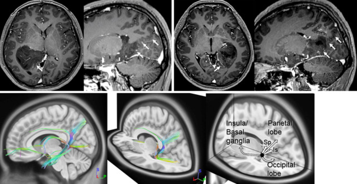
Credit: Figure used with permission from Shibahara et al.
Charlottesville, VA (December 14, 2021). Researchers from four academic neurosurgical centers in Japan found that the parietooccipital fissure, which divides the temporal, occipital, and parietal lobes of the brain, acts as an obstacle that may result in less invasion of posterior medial temporal gliomas toward the occipital lobe.
Detailed findings, including images obtained with magnetic resonance and diffusion tensor imaging and fiber tractography, are provided in the article “Role of the parietooccipital fissure and its implications in the pathophysiology of posterior medial temporal gliomas,” by Ichiyo Shibahara, MD, PhD, and colleagues, published today in the Journal of Neurosurgery (https://thejns.org/doi/10.3171/2021.7.JNS21990).
Gliomas are the most common primary brain tumors and have a poor prognosis even with currently available therapies. An important treatment strategy is surgical removal of as much tumor as possible without damaging important functional areas of the brain, a precaution that requires detailed information regarding the individual anatomy of each patient.
In this study, the authors performed tumor removal and assessed the anatomical architecture between the tumor location and the parietooccipital fissure in 24 patients with posterior medial temporal gliomas. In all patients, the anterior end of the parietooccipital fissure was the posterior border of the tumor. Magnetic resonance imaging of 20 patients showed that the tumor invaded along and anterior to the parietooccipital fissure, extending to the isthmus of the cingulate gyrus and the splenium of the corpus callosum. Fiber tracts running through the posterior medial temporal lobe were observed to correspond to these invasion patterns. Diffusion tensor imaging and fiber tractography also showed that fibers running toward the occipital lobe made a detour to avoid the parietooccipital fissure. These findings highlight the importance of the parietooccipital fissure in determining the invasion pattern of posterior medial temporal gliomas.
As the researchers state, their evidence confirms that in patients with posterior medial temporal glioma the parietooccipital fissure is an uninterrupted main sulcus comprising the posterior boundary of glioma invasion. Therefore, this fissure is a key landmark for understanding the complex anatomical architecture and invasion pattern of posterior medial temporal gliomas during preoperative analysis, information that may lead to more effective surgical treatment.
When asked about the study, Dr. Shibahara said, “All neurosurgeons/neuro-oncologists share some impression that gliomas invade in a specific direction; however, only a few studies have focused on demonstrating such phenomena. In this study we noticed that posterior medial temporal gliomas in different patients presented not only similar invasion patterns but also radiological features indicating a clear posterior border. We found that invasion of glioma cells was highly influenced by the parietooccipital fissure, suggesting a generalized pattern in which sulci act as the anatomical boundary of glioma invasion.”
Shibahara I, Saito R, Kanamori M, Sonoda Y, Sato S, Hide T, Tominaga T, Kumabe T. Role of the parietooccipital fissure and its implications in the pathophysiology of posterior medial temporal gliomas. Journal of Neurosurgery, published online, ahead of print, December 14, 2021; DOI: 10.3171/2021.7.JNS21990.
Disclosure: The authors report no conflict of interest concerning the materials or methods used in this study or the findings specified in this paper.
Funding: Supported in part by the Japan Society for the Promotion of Science KAKENHI for Early-Career Scientists, grant number 18K16569; a Research Grant for young medical doctors and healthcare professionals from SRL, Inc.; and grants from the Ichiro Kanehara Foundation, the Yokoyama Rinsho Yakuri Foundation, the Akaeda Igaku Kenkyu Foundation, and the Uehara Memorial Foundation. Data were provided (in part) by the Human Connectome Project, WU-Minn Consortium (Principal Investigators: David Van Essen and Kamil Ugurbil; 1U54MH091657), funded by the 16 NIH Institutes and Centers that support the NIH Blueprint for Neuroscience Research and by the McDonnell Center for Systems Neuroscience at Washington University.
###
For additional information, please contact: Gillian Shasby, Director of Publications, Journal of Neurosurgery Publishing Group, One Morton Drive, Suite 200, Charlottesville, VA 22903. Email: [email protected]; Phone: 434-924-5555.
For 77 years, the Journal of Neurosurgery has been recognized by neurosurgeons and other medical specialists the world over for its authoritative clinical articles, cutting-edge laboratory research papers, renowned case reports, expert technical notes, and more. Each article is rigorously peer reviewed. The Journal of Neurosurgery is published monthly by the JNS Publishing Group, the scholarly journal division of the American Association of Neurological Surgeons. Other peer-reviewed journals published by the JNS Publishing Group each month include Neurosurgical Focus, the Journal of Neurosurgery: Spine, and the Journal of Neurosurgery: Pediatrics. All four journals can be accessed at www.thejns.org.
Founded in 1931 as the Harvey Cushing Society, the American Association of Neurological Surgeons (AANS) is a scientific and educational association with more than 12,000 members worldwide. Fellows of the AANS are board-certified by the American Board of Neurological Surgery, the Royal College of Physicians and Surgeons of Canada, or the Mexican Council of Neurological Surgery, A.C. Neurosurgery is the medical specialty concerned with the prevention, diagnosis, treatment and rehabilitation of disorders that affect the spinal column, spinal cord, brain, nervous system and peripheral nerves. For more information, visit www.AANS.org.
Journal
Journal of Neurosurgery
DOI
10.3171/2021.7.JNS21990
Method of Research
Observational study
Subject of Research
People
Article Title
Role of the parietooccipital fissure and its implications in the pathophysiology of posterior medial temporal gliomas
Article Publication Date
14-Dec-2021
COI Statement
The authors report no conflict of interest concerning the materials or methods used in this study or the findings specified in this paper.
