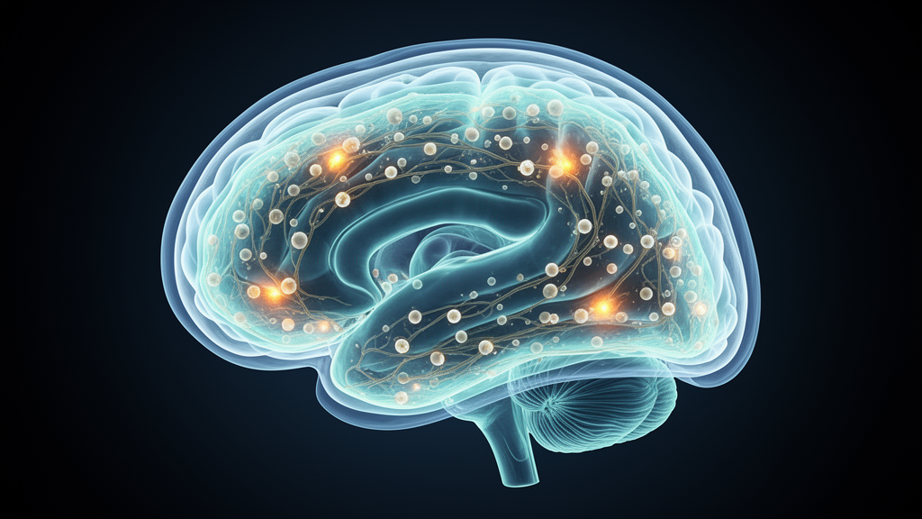
Deep beneath the skull, three delicate membranes known as the meninges wrap the brain and spinal cord in a living shield. Long treated as little more than passive packaging for the central nervous system, these layers are now recognized as dynamic hubs of immune surveillance, vascular regulation and barrier defense. A new study published in Nature Cell Biology has delivered the most comprehensive picture yet of how this remarkable tissue assembles itself during human development, charting the birth of its cellular residents from the sixth to the twenty-first week of pregnancy and, in a striking twist, revealing how a single extra chromosome reshapes the entire landscape.
An international team led by researchers at the University of Cambridge and the Wellcome Sanger Institute, working under the direction of Sarah Teichmann, constructed a paired atlas of single-nucleus RNA sequencing and single-nucleus assay for transposase-accessible chromatin sequencing, capturing both the gene expression and the open chromatin landscape of individual meningeal cells. To anchor these molecular profiles in physical space, the team added highly multiplexed spatial transcriptomics using a custom 310-plex Xenium panel, alongside in situ sequencing and multiplexed protein imaging. The samples came from seventeen donors spanning six to twenty-one postconception weeks, drawn from both anterior and posterior cranial regions, and included three samples with trisomy 21, the chromosomal cause of Down syndrome.
The resulting atlas identified fifty-one transcriptional clusters encompassing roughly 150,000 nuclei, dominated by fibroblasts and endothelial cells but also containing a full repertoire of immune populations, including B cells, T cells, myeloid cells, erythroid cells, eosinophils, basophils and mast cells. Myeloid and erythroid states were enriched in the first trimester, while lymphoid populations expanded from the beginning of the second trimester, mirroring patterns of lymphoid maturation seen across other fetal tissues. The depth of the dataset allowed the researchers to resolve fine-grained subtypes within each compartment and to trace how their proportions shifted across developmental time.
Among the most technically significant findings concerns the blood-brain barrier, the specialized endothelial lining that strictly gates which molecules can enter the brain. The team captured paired RNA and chromatin profiles of developing barrier-forming capillary endothelial cells and mapped them along an arteriovenous axis, identifying arterial cells marked by GJA5, venous cells marked by EMCN and TLL1, and barrier capillary cells enriched for the transcription factor RGCC. Applying the SCENIC+ framework to predict gene regulatory networks, the researchers found that the transcription factor ETS1 sits at the center of a network directly controlling solute carrier genes, including SLC7A5, SLC7A8 and SLC43A2, which encode amino acid transporters. SLC7A5 is known to mediate uptake of l-DOPA, the precursor of the dopamine that is depleted in Parkinson’s disease, underscoring how developmental wiring of the barrier shapes drug delivery decades later. Additional barrier-specific regulators included FOXF2, FOXQ1 and DEAF1, the last of which has been linked to neurodevelopmental delay, hinting at a molecular coupling between barrier formation and brain development itself.
The fibroblast compartment yielded an equally compelling developmental story. Subclustering revealed seven meningeal fibroblast states whose marker genes correspond to the pial, arachnoid and dural layers described in mouse studies: pial fibroblasts expressed LAMA2 and LAMA1, arachnoid fibroblasts expressed SLC7A2 and CRABP2, and dural fibroblasts expressed MGP, MATN4 and NPPC. Pseudotime modeling and differential abundance testing indicated that pial fibroblast states act as early progenitors, enriched in the first trimester, from which arachnoid and dural fibroblasts emerge later. All fibroblast clusters expressed HIC1, a marker of embryonic mesenchymal progenitors whose knockout in mice abolishes dura formation and causes severe cranial defects. Spatial mapping at single-cell resolution confirmed that these molecular states occupy anatomically distinct layers, and because the dura and leptomeninges are not yet separable at six weeks but are clearly layered by nine weeks, the data suggest that the meningeal layers physically partition within the first trimester. Gene set enrichment of anterior-region fibroblasts pointed to neural crest differentiation as the top pathway, consistent with the idea that anterior meningeal fibroblasts derive from cranial neural crest while posterior populations arise from mesoderm.
The immune findings may prove the most consequential. The atlas captured macrophage progenitors, microglia-like clusters and three border-associated macrophage populations, one of which, marked by low FOLR2 expression, proved to be intimately associated with blood vessels. The progenitor cluster expressed RAN and STMN1, signatures of yolk sac-derived macrophage precursors, and computational integration with published yolk sac and fetal liver atlases showed that meningeal macrophage progenitors and microglia sit transcriptionally closer to yolk sac than to liver progenitors. The data therefore support a model in which yolk sac-derived precursors seed the meninges and continuously give rise to both microglia and border-associated macrophages through the late second trimester. CellPhoneDB interaction analysis predicted that the perivascular macrophages send pro-angiogenic signals, including TGFB1 and IGF1, to endothelial cells, while endothelial cells reciprocate with GAS6, a macrophage recruitment factor, suggesting a self-reinforcing loop in which immune cells are drawn to vessels and then promote their growth. These cells also expressed class II MHC strongly, raising the possibility that they serve as immune sentinels monitoring intravascular pathogens after birth.
Perhaps the most surprising discovery was the extent of B cell development within the prenatal meninges. The atlas captured essentially the entire B lineage, from early progenitors through precursor, immature and mature plasma cell states, and differential abundance testing showed B lineage enrichment rising sharply in the second trimester. To explain how these cells arrive, the team built a new computational tool called cell2home, which infers chemotactic homing by aggregating chemokine receptor-ligand signaling into a single migration-oriented affinity score between cell types. Cell2home predicted that early B progenitors are attracted into blood vessels through CD34/PODXL ligands engaging the receptor SELL on the B cells, while more mature B states are drawn into tissue by the chemokine CCL19 from dural fibroblasts acting on CCR7, with a developmental switch from SELL to CCR7 expression underlying the transition. Spatial analysis independently confirmed that progenitor B cells colocalize with vasculature, and protein staining revealed CD20-positive B cells within both the cranial bone marrow and the dural meninges at sixteen weeks, a physical association between developing B cells and the meningeal niche that had never been described in humans.
The trisomy 21 samples added a disease dimension to the atlas. After age-matched integration with euploid samples, the team observed a clear gene dosage effect, with excess differentially expressed genes mapping to chromosome 21. Stromal cells, particularly the early pial fibroblast progenitors, were depleted in trisomic samples, and fibroblast chemokine expression was markedly reduced: CXCL1, CXCL2, CXCL3 and CXCL8, all targets of NF-kB signaling, were downregulated alongside the MAPK and NF-kB pathways themselves. In contrast, the immune compartment showed enhanced JAK-STAT signaling and upregulation of interferon-stimulated genes including IFI44, MX2, OAS, ISG15 and MX1, consistent with the interferon hyperactivation long associated with Down syndrome. Enrichment of KRAS and MTORC1 signaling further echoed molecular programs implicated in trisomy 21-associated leukemia and metabolic phenotypes, though the authors caution that functional experiments and larger, stage- and sex-matched cohorts are needed to confirm these disease mechanisms.
Alongside cell2home, the team released a second analytical tool, Nichefinder, which detects spatial niches by combining cell-type label transfer, spatial nearest-neighbor aggregation and non-negative matrix factorization to identify recurring motifs of colocalizing cell types. Benchmarking against the existing NiCO method on simulated data showed near-identical interaction detection, with correlations of 0.86 and 0.82 across two scenarios, while comparisons with NicheCompass on mouse hippocampus data showed that Nichefinder excels at resolving dispersed cell types such as astrocytes, microglia and endothelial cells rather than broad contiguous domains. Both tools are openly available, and the authors envision cell2home being applied to immune recruitment in inflammation and cancer as well as development.
The study leaves open questions that will drive the next wave of work. The neural crest progenitors of the anterior meninges were not captured, and profiling stages before five weeks will be needed to characterize them. Direct lineage tracing of the proposed yolk sac origin of meningeal macrophages remains technically formidable in human embryos, and the postnatal fate of meningeal-resident B cells, including whether bone marrow and meningeal clones share progenitors, could be probed with B cell receptor sequencing. Nevertheless, by defining the cellular and regulatory programs that assemble the meninges and establish early barrier immunity, this atlas provides an essential reference for understanding congenital neurodevelopmental disease, meningioma origins and the immune defenses that guard the brain for a lifetime.
Subject of Research: Single-cell and spatial multiomic mapping of human meningeal development from 6 to 21 postconception weeks, including blood-brain barrier endothelium, myeloid and B cell lineages, and trisomy 21 effects
Article Title: Spatially resolved multiomics of human meningeal development reveal lineage and disease dynamics
Article References: To, K., Pett, J. P., Dufva, O., Chipampe, N.-J., Polanski, K., Cujba, A.-M., Roberts, K., Predeus, A. V., Yayon, N., Kanemaru, K., Patel, M. I., Horsfall, D., Li, T., Mazin, P., Cakir, B., Kapuge, R., Wilk, A., Memi, F., He, X., … Teichmann, S. A. (2026). Spatially resolved multiomics of human meningeal development reveal lineage and disease dynamics. Nature Cell Biology. https://doi.org/10.1038/s41556-026-02075-8
Image Credits: AI Generated
DOI: 10.1038/s41556-026-02075-8
Keywords: meninges, single-nucleus multiomics, spatial transcriptomics, blood-brain barrier, yolk sac macrophages, B cell development, fibroblast lineages, trisomy 21, gene regulatory networks, cell2home, Nichefinder, fetal neuroimmunology
News Source: Cassandra Pierce. (October 9, 2026). First Multiomic Atlas Reveals How the Human Brain’s Protective Layers Build Their Immune Defenses. Scienmag.