Blood tests can reveal that cancer is present and characterize tumor DNA, cells, or molecular alterations, but they generally cannot show where metastatic disease has developed. A proposed framework called Spatial Liquid Biopsy would use extracellular vesicles released by tumors to infer the anatomical sites of metastases, potentially extending liquid biopsy from molecular detection to spatial mapping. The concept, described in a 2026 Frontiers in Oncology perspective, remains a hypothesis: direct evidence that circulating tumor-associated vesicles carry surface proteins identifying the organ containing an established metastasis is still limited, and no clinical assay has yet been validated.
Metastasis accounts for approximately 90% of cancer-related deaths, according to the perspective, making the distribution of secondary tumors a central problem in oncology. Physicians currently combine CT, MRI, PET, pathological examination, serum markers, and blood-based analyses to assess metastatic disease. Imaging can map lesions anatomically, while liquid biopsies can track tumor burden, molecular changes, treatment response, and resistance. The proposed Spatial Liquid Biopsy approach would seek a missing layer of information: the probable location of disease inferred from a peripheral blood sample, rather than from an anatomical image.
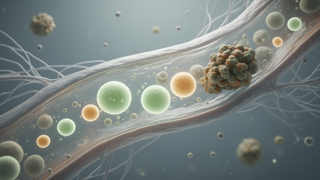
Extracellular vesicles, or EVs, are membrane-bounded particles released by cells that carry proteins, lipids, and nucleic acids. Their molecular composition often reflects that of the cells that produced them, and they can participate in communication between cancer cells and their surroundings. Tumor-associated proteins on EV membranes can be used to enrich a population of vesicles from blood. Candidate capture markers discussed in the article include EpCAM, MUC1, and EGFR for lung cancer; EpCAM, HER2, and MUC1 for breast cancer; and EpCAM and CEA for colorectal cancer. Such markers are not exclusive to cancer, however, because normal epithelial cells can also express them, complicating the separation of tumor-associated EVs from the much larger background of vesicles released by healthy tissues.
Spatial Liquid Biopsy is based on a two-stage analytical principle. First, tumor-associated EVs would be captured with a selected surface marker or marker combination. Those vesicles would then be examined for extracellular proteins associated with particular organs. Candidate organ-associated proteins include brevican and neurocan for the brain, leukocyte cell-derived chemotaxin-2 and secreted phosphoprotein 2 for the liver, alkaline phosphatase and bone sialoprotein for bone, and surfactant-associated proteins A1 and C for lung. The goal would be a double-positive population bearing both tumor-associated and organ-associated signals, which the framework proposes could be more spatially specific than either dimension alone.
The biological hypothesis is that cancer cells colonizing another organ interact with local stromal, immune, endothelial, extracellular-matrix, and tissue-resident cells. In the liver, those interactions involve cell types such as Kupffer cells and hepatic stellate cells; in the brain, astrocytes and microglia; and in bone, osteoblasts, osteoclasts, and marrow stromal cells. These exchanges may influence both the metastatic tumor and the vesicles it releases. Organ-associated extracellular proteins might become associated with the surface of locally generated tumor EVs, allowing the vesicles to carry a composite signature of malignancy and location. This mechanism has not yet been directly demonstrated for the proposed strategy, making it a central question for future experiments.
Existing studies provide indirect support. Experimental work has shown that tumor-derived exosomes from models with different metastatic organ preferences carried distinct surface integrin profiles: α6β4 and α6β1 were associated with lung tropism, while αvβ5 was associated with liver tropism. Those integrins are tumor-cell proteins that help determine where cancer cells travel, rather than proteins derived from the destination organ, so they do not prove the Spatial Liquid Biopsy mechanism. Nevertheless, they demonstrate that the surface composition of tumor EVs can contain information linked to organ-specific metastatic behavior.
Human evidence comes from a study comparing plasma EV proteomes among patients with lung cancer and either solitary liver metastasis, solitary brain metastasis, or locally advanced disease without distant metastasis. The investigators found EV-associated protein profiles that differed by metastatic site and subsequently validated candidate proteins associated with brain or liver metastases in a larger cohort. Because the analysis examined bulk plasma EVs rather than EVs specifically enriched for tumor origin, the cellular source of the detected proteins could not be assigned to cancer cells, metastatic organs, or other host cells. The results nevertheless suggest that circulating EV protein profiles can carry information associated with the anatomical site of established disease.
Separate cell-culture experiments found that breast cancer cell lines with different organotropic metastatic phenotypes released EVs with distinct proteomic profiles. That supports an association between metastatic phenotype and EV composition, but the vesicles were collected from cultured tumor cells rather than from animals or patients with established metastases. Taken together, the evidence supports three observations: tumor EVs can carry proteins linked to metastatic tropism, bulk circulating EV profiles can differ according to metastatic site in patients, and tumor cells with different organotropic phenotypes can release compositionally distinct EVs. It does not yet establish that blood-borne, tumor-enriched EVs display organ-derived surface proteins in the manner required by the new framework.
If validated, multiplex profiling could reveal several metastatic locations in one blood sample. A patient with liver and bone metastases might have separate EV subpopulations associated with each organ, rather than individual vesicles carrying markers from both sites. The absolute abundance of a spatial signal should not automatically be read as a direct measure of metastatic burden. EV levels can also be influenced by tumor volume, release rates, vascular access, clearance, treatment effects, and the biology of individual metastatic microenvironments. Determining whether a signal tracks lesion number or volume would therefore require comparison with independently established disease burden, while repeated measurements within a patient could test whether changes parallel progression or regression at particular sites.
Development would require staged validation. An initial proof-of-concept study could test the proposed marker panel in archived plasma from patients with anatomically confirmed metastases, using single-dominant-site cohorts to ask whether the signatures distinguish brain, bone, liver, or other locations. Lung cancer could serve as an initial model because it commonly spreads to several anatomically distinct organs. Retrospective validation would need to precede studies involving multiorgan disease, and prospective longitudinal studies would have to determine whether emerging signals precede radiographic detection and whether changes correspond to pathology, treatment response, and clinical outcomes. Improvements in EV enrichment, antibody engineering, multiplex protein detection, ultrasensitive assays, single-EV analysis, and computational interpretation may eventually improve performance, but the proposed starting markers must first be tested directly.
The perspective positions Spatial Liquid Biopsy as a possible complement to imaging rather than a replacement. Its ultimate value would depend on whether prospective studies show that blood-based organ signals can reliably identify metastatic involvement, reveal changes earlier than current surveillance methods, or support more targeted use of diagnostic imaging. The framework is scientifically testable, but its strongest claims remain conditional: it has not yet been shown that organ-associated proteins bind to circulating tumor EVs, that such double-positive vesicles accurately locate metastases, or that their abundance reflects tumor burden. If those links are established, the approach could turn extracellular vesicles into minimally invasive molecular maps of where cancer has spread; without them, it would remain a promising but unvalidated strategy for expanding liquid biopsy beyond answering whether cancer is present to addressing where metastatic disease may be located.
Subject of Research: Conceptual use of extracellular-vesicle biomarkers to infer the anatomical location of cancer metastases from blood
Article Title: From liquid biopsy to spatial liquid biopsy: extracellular vesicles as molecular maps of cancer metastasis
Article References: Mitsuhashi, M. (2026). From liquid biopsy to spatial liquid biopsy: extracellular vesicles as molecular maps of cancer metastasis. Frontiers in Oncology, 16, Article 1940169. https://doi.org/10.3389/fonc.2026.1940169
Image Credits: AI Generated
DOI: 10.3389/fonc.2026.1940169
Keywords: liquid biopsy, cancer metastasis, extracellular vesicles, spatial biomarkers, blood biomarkers, tumor microenvironment, precision oncology, organ-specific metastasis, cancer surveillance, liquid, biopsy, spatial
News Source: Nathaniel Bowman. (October 21, 2026). Extracellular Vesicles Could Map the Location of Metastatic Cancer. Scienmag.