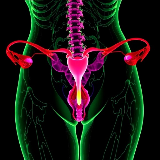

Recent advances in cellular and molecular technologies have ushered in a transformative era in the study of endometriosis, a complex and enigmatic hormone-sensitive disorder affecting millions worldwide. Utilizing cutting-edge tools like single-cell RNA sequencing and mass spectrometry, researchers are uncovering unprecedented insights into the heterogeneous nature of endometriotic lesions and the eutopic endometrium. These technologies illuminate diverse cell populations, unique molecular signatures, and intricate cellular interactions, all of which deepen our understanding of the pathophysiological underpinnings of the disease. The integration of these datasets promises to untangle the complex biology behind varying lesion subtypes and the manifestation of symptoms, advancing towards personalized diagnostic and therapeutic strategies.
A major breakthrough lies in the spatial resolution of transcriptomic data, enabling scientists not only to catalog distinct cell types but also to map their precise locations within the affected tissues. This spatial context is critical, as the microenvironment and proximity of lesions appear to influence disease progression and symptomatology. Endometriotic lesions, presenting with variable histological features such as differing proportions of glandular epithelium and stromal fibroblasts, challenge traditional diagnostic paradigms. This cellular heterogeneity, combined with lesion fibrosis, hemosiderin deposits, and metaplastic changes, complicates pathological confirmation. Recent efforts to standardize lesion documentation, supported by surgeon collaboration, highlight the importance of lesion localization and tissue architecture in guiding meaningful analysis.
The adoption of standard operating procedures (SOPs) in the collection, processing, and storage of biospecimens also represents a crucial stride toward reproducibility and data harmonization. The World Endometriosis Research Foundation’s Endometriosis Phenome and Biobanking Harmonization Project (WERF-EPHeCT) has spearheaded protocols ensuring uniform handling of tissues and peripheral blood, which are essential for high-fidelity molecular analyses. These rigorously maintained SOPs facilitate cross-study comparisons and meta-analyses that are vital for deciphering the complexities of endometriosis. Furthermore, leveraging both frozen and formalin-fixed samples allows investigations ranging from transcriptomic to proteomic and metabolomic profiling, broadening the scope of molecular insights.
Understanding the hormonal milieu at the time of sample acquisition is paramount, as fluctuating estrogen and progesterone levels profoundly impact tissue transcriptomes and cellular phenotypes. Quantification of circulating estradiol and progesterone, coupled with histologic menstrual phase dating of endometrial samples, empowers researchers to contextualize molecular data within precise hormonal stages. Importantly, the field is beginning to appreciate the nuanced distinctions between endogenous ovarian hormones and exogenous hormonal therapies such as progestins, which activate overlapping but distinct signaling pathways. This recognition underscores the necessity to meticulously document patients’ hormonal treatments, thereby refining interpretations of molecular findings and responses to therapy.
The meticulous definition of cases and controls in endometriosis research remains a cornerstone for the validity and translation of findings. Clinical metadata encompassing pain characteristics, fertility status, comorbid gynecologic diseases, and demographic factors are foundational for stratifying study participants. Platforms like REDCap enable ongoing, dynamic documentation of these covariates, facilitating robust correlation between clinical phenotypes and molecular data. However, current limitations include underrepresentation of diverse populations and insufficient ethnic documentation, which restricts the generalizability of results. Addressing cohort diversity emerges as a pressing opportunity to enrich the understanding of endometriosis across different genetic and environmental contexts.
Histopathological confirmation of lesions presents additional challenges due to the variable presence of key cellular components. While approximately 60–80% of biopsied lesions demonstrate definitive endometrial-like epithelium and stromal fibroblasts, termed “stromal endometriosis” can lack epithelial elements, complicating diagnosis. Immunohistochemical staining, especially for CD10—a marker of endometrial stromal cells—has become a vital adjunctive tool in these ambiguous cases. Moreover, cutting-edge computer-aided histopathology is beginning to revolutionize lesion identification with enhanced objectivity and accuracy, suggesting a future where artificial intelligence may aid routine diagnosis.
The small size and fibrotic nature of certain lesion types, such as superficial peritoneal endometriosis (SPE) and deep infiltrating endometriosis (DIE), pose particular difficulties for molecular studies. Limited tissue yields and low viable cell counts can restrict the depth of single-cell analyses and proteomic quantification. These physical constraints necessitate innovative sampling methods and sensitive analytical platforms capable of generating comprehensive data from minimal inputs. The field is actively pursuing such technological innovations to overcome these barriers and ensure robust characterization of all lesion subtypes.
Advancements in untargeted proteomics and metabolomics using mass spectrometry are expanding the molecular landscape beyond nucleic acids to include proteins and small molecules. The ability to quantify thousands of proteins and metabolites simultaneously in blood and tissue specimens opens new avenues to identify biomarkers and elucidate biochemical pathways perturbed in endometriosis. These multi-omic approaches promise to capture the dynamic interplay between genomic, proteomic, and metabolic alterations, providing a holistic picture of disease biology and its systemic effects, including common comorbidities.
Given the disease’s hormone-sensitivity, it is critical to interpret multi-omic data within the hormonal context. Variations in estrogen and progesterone influence gene expression, immune responses, and tissue remodeling, which can differ between lesion locations and phases of the menstrual cycle. Consequently, phase-specific transcriptomic signatures must be integrated into analytical frameworks to avoid confounding hormonal influences with disease-specific alterations. Future investigations will likely refine these signatures further, improving the specificity of molecular diagnostics and informing targeted therapeutic interventions.
Data heterogeneity resulting from differences in study designs, methodologies, and sample processing protocols continues to limit the reproducibility and clinical translation of research findings. Addressing this challenge depends on concerted community efforts to standardize experimental workflows and data reporting. The WERF-EPHeCT consortium’s protocols embody these principles and serve as a global reference to foster rigor. Additionally, advanced bioinformatic approaches that account for variability and integrate heterogeneous datasets will be essential to harness the full potential of multi-omic data and bridge the gap between bench and bedside.
The deployment of artificial intelligence and machine learning algorithms is emerging as a powerful strategy to disentangle complex biological patterns inherent in endometriosis. These tools can manage vast, multidimensional datasets, uncover subtle molecular signatures, and predict disease trajectories or treatment responses. When combined with comprehensive clinical phenotyping and robust biobanking infrastructure, AI-driven analyses hold promise to revolutionize diagnostics, patient stratification, and personalized management plans, ultimately improving outcomes for affected individuals.
Importantly, the disease’s association with a spectrum of gynecologic comorbidities—in particular uterine fibroids, adenomyosis, and abnormal uterine bleeding—complicates clinical discernment and molecular analyses. The co-occurrence of these disorders in both cases and controls necessitates nuanced phenotypic characterization to isolate disease-specific signatures. Comprehensive metadata capturing these overlapping conditions is thus indispensable. Future research integrating multi-omic data with detailed clinical annotations may shed light on shared or distinct pathological mechanisms, potentially unveiling novel therapeutic targets.
Endometriosis research is also expanding beyond lesion analysis to include circulating biomarkers in peripheral blood, facilitating non-invasive diagnostic approaches. Molecular signatures detectable in plasma or serum could revolutionize early detection and monitoring, mitigating current reliance on invasive laparoscopic diagnosis. Mass spectrometry and transcriptomic profiling of blood components stand at the forefront of this endeavor, with preliminary studies indicating promising candidate markers awaiting validation in larger, diverse cohorts.
Finally, the implications of these advancements extend to therapeutic innovation. A molecularly informed understanding of endometriosis pathogenesis paves the way for precision medicine approaches—tailoring treatments based on lesion subtype, hormonal milieu, and molecular profiles. As hormonal therapies display differential pathway activations depending on chemical composition and patient biology, future interventions may optimize efficacy while minimizing side effects. This paradigm shift promises to improve quality of life and reproductive outcomes for those affected.
In conclusion, the convergence of single-cell technologies, spatial transcriptomics, proteomics, metabolomics, and artificial intelligence is reshaping the landscape of endometriosis research. Despite lingering challenges inherent to sample heterogeneity, technical variability, and disease complexity, the trajectory is unmistakably toward more refined, mechanistic insights and clinically impactful discoveries. The collaborative infrastructure, exemplified by global biobanking and phenotyping initiatives, underpins this progress. With continued integration of diverse populations and comprehensive clinical metadata, the field is poised to close knowledge gaps and translate multi-omic findings into personalized diagnostics and therapeutics for this pervasive and debilitating disease.
Subject of Research: Endometriosis disease phenotyping using advanced molecular technologies combined with clinical data and artificial intelligence.
Article Title: Real world perspectives on endometriosis disease phenotyping through surgery, omics, health data, and artificial intelligence.
Article References:
Nezhat, C.R., Oskotsky, T.T., Robinson, J.F. et al. Real world perspectives on endometriosis disease phenotyping through surgery, omics, health data, and artificial intelligence. npj Womens Health 3, 8 (2025). https://doi.org/10.1038/s44294-024-00052-w
Image Credits: AI Generated
Tags: advances in endometriosis researchAI insights in endometriosis treatmentcellular interactions in endometriosischallenges in endometriosis diagnosisendometriosis phenotyping advancesheterogeneous nature of endometriosismass spectrometry for endometriosis researchmolecular signatures of endometriotic lesionspathology of endometriotic lesionspersonalized diagnostic strategies for endometriosissingle-cell RNA sequencing in endometriosisspatial transcriptomics in endometriosis

