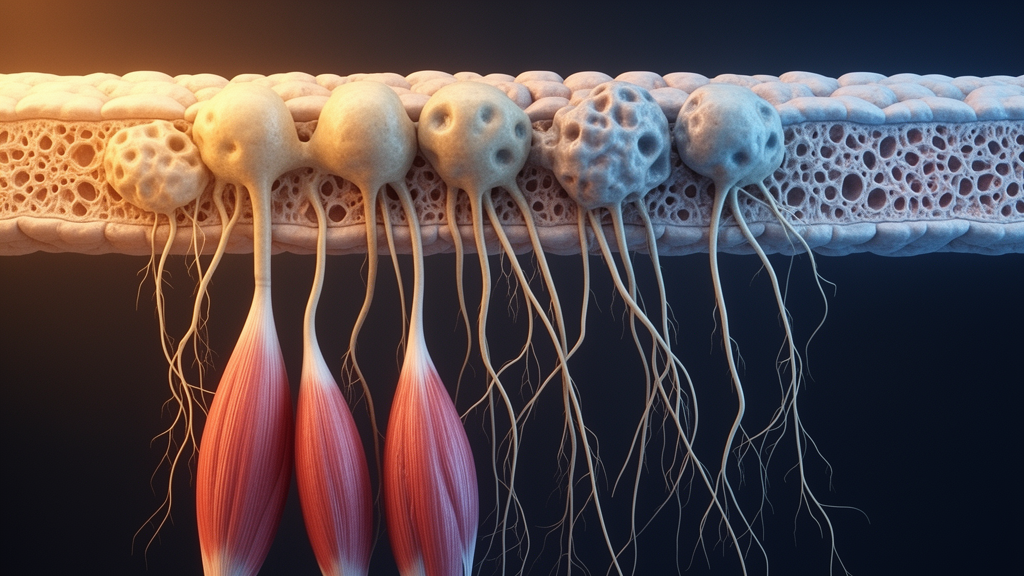
A form of muscular dystrophy long defined by the wasting of limb and girdle muscles may actually announce itself much higher up the body. A new study combining patient MRI scans with longitudinal imaging in mice reports that the muscles flanking the spine degenerate early, progressively, and in some cases before the limb muscles that give the disease its name. The work, published in EMBO Molecular Medicine, also identifies the cellular and molecular machinery behind this fatty replacement and points to an enzyme already targeted by experimental drugs as a possible way to stop it.
Dysferlinopathy, classified as limb girdle muscular dystrophy R2, is an inherited disorder caused by mutations in the DYSF gene, which reduce or eliminate the muscle protein dysferlin. Dysferlin normally sits in the membrane surrounding muscle fibers and enables calcium-dependent repair of membrane injuries; without it, damaged fibers fail to heal, die, and are progressively replaced by fat and connective tissue, typically beginning in early adulthood. No approved therapy currently exists for the disease, and anti-inflammatory corticosteroids have proven ineffective despite chronic muscle inflammation being a feature of the condition.
To investigate the role of spinal muscles, the researchers retrospectively analyzed 20 dysferlinopathy patients diagnosed at hospitals in South Korea and France, all with biallelic disease-causing mutations confirmed by genetic sequencing and all suffering progressive limb-girdle weakness with elevated creatine kinase levels. On T1-weighted MRI scans, the patients showed high signal intensity in the erector spinae muscles of the lower back, indicating fatty replacement, while adjacent pelvic girdle muscles such as the psoas and quadratus lumborum, along with limb muscles, remained largely unaffected. Within the erector spinae group, the iliocostalis muscle was the earliest and most severely affected, followed by the longissimus and then the spinalis, and the lower back was more involved than the upper back and neck regions.
The severity of spinal involvement also tracked with disease activity: higher initial serum creatine kinase levels, a marker of ongoing muscle breakdown, were associated with an earlier age of disease onset and an earlier age at which spinal MRI was required. Because fatty replacement seen on scans in patients could theoretically be a secondary consequence of limb weakness forcing the spine to compensate during upright walking, the team turned to a quadruped animal model, the BLA/J mouse, which carries a dysferlin mutation and develops limb muscle pathology closely resembling the human disease.
Longitudinal MRI of BLA/J mice at 6, 8, 10, and 22 months of age revealed that fatty degeneration appears first in the spinal muscles, detectable by 6 months of age, when the quadriceps show little to no fatty change. Quadriceps degeneration only became MRI-detectable at around 10 months and progressed thereafter. At the symptomatic age of 10 months, spinal muscles of BLA/J mice showed roughly 25-fold greater fatty replacement compared with wild-type controls, versus about fivefold greater degeneration in quadriceps. Wild-type mice showed minimal degeneration even at 22 months. The authors argue this ordering shows spinal muscle loss is a primary feature of dysferlin deficiency rather than a byproduct of bipedal posture or limb weakness.
The spinal degeneration also had functional consequences. Symptomatic BLA/J mice lost the natural curvature of the spine, adopting a visibly flattened posture, and treadmill gait analysis showed that on both 15-degree inclines and declines the mice walked with longer strides, longer stride and stance durations, and reduced forelimb and hindlimb stride frequency at a fixed treadmill speed. On horizontal treadmills, their gait parameters were largely normal, suggesting the animals compensate on slopes by taking fewer but longer and slower steps. The authors suggest that recognizing similar compensatory strategies in patients could inform rehabilitation approaches aimed at delaying mobility decline.
Histological examination traced the cellular sequence underlying the imaging findings. As early as 6 months of age, BLA/J spinal muscles showed centrally nucleated regenerating fibers, immune cell infiltration, and fibrofatty degeneration, which worsened progressively. Immunostaining revealed that damaged, IgM-positive myofibers accumulated first in the iliocostalis, and these areas were simultaneously enriched in fibroadipogenic progenitors, or FAPs, marked by the protein PDGFRα. Over time, the damaged fibers were replaced by perilipin-labeled adipocytes. FAPs isolated from the iliocostalis of BLA/J mice also showed significantly greater spontaneous adipogenic differentiation in culture than FAPs from wild-type iliocostalis or from BLA/J quadriceps, indicating that the degenerating spinal muscle environment intrinsically biases these progenitors toward a fat-producing fate.
To find druggable controls for this shift, the team performed RNA sequencing on FAPs from 10-month-old BLA/J and wild-type mice, identifying 1,035 differentially expressed genes and implicating HDAC and Wnt/β-catenin signaling as the principal regulatory pathways. Pharmacological testing in cultured spinal FAPs showed that the HDAC inhibitor givinostat, recently approved for Duchenne muscular dystrophy, and the matrix metalloproteinase inhibitor batimastat each partially suppressed spontaneous adipogenesis. The GSK3 inhibitor LY2090314, which acts on the Wnt/β-catenin pathway, fully abolished adipogenic differentiation in vitro. Mechanistically, dysferlinopathic FAPs showed elevated PPARγ, reduced active β-catenin, and increased activated GSK3β; LY2090314 reduced the activated form of GSK3β, restored β-catenin signaling, and lowered PPARγ expression.
In vivo testing supported the target’s promise but also flagged safety concerns. Treating 7-month-old BLA/J mice with LY2090314 on alternate days for six weeks notably preserved spinal muscle and reduced perilipin-marked fatty replacement, and the drug completely protected tibialis anterior muscles from fat accumulation after three rounds of experimentally induced injury. However, treated mice developed adverse effects, including scarring of the skin and diaphragm at the injection site and enlargement of the spleen, which the authors say must be weighed in any effort to develop long-term therapy. They note that alternative GSK3 inhibitors, such as tideglusib, which has reached late-stage clinical testing for myotonic dystrophy, may offer safer routes.
The study carries important limitations. The human component was a retrospective, cross-sectional cohort of 20 patients, which can show associations between MRI findings and disease markers but cannot establish when spinal degeneration begins in people; the temporal evidence for early onset comes from the mouse model, with small groups of three to ten animals per analysis. The drug findings are preclinical, obtained in a mouse model and in cultured cells, and cannot be extrapolated directly to patient benefit. Even so, by establishing fatty degeneration of spinal muscles as an early, conserved hallmark of dysferlinopathy in both species and identifying FAP adipogenesis driven through GSK3 signaling as its mechanism, the work reframes the disease as an axial as well as limb disorder and opens a concrete therapeutic avenue for a condition that currently has none.
Subject of Research: Early fatty degeneration of spinal muscles in dysferlinopathy and its mechanism in fibroadipogenic progenitors
Article Title: FAP adipogenesis drives early spinal muscle loss in dysferlinopathy and reveals a therapeutic target
Article References: Moon, Y. J., McCormack, N., Diehn, T., Lin, S., Staedler, K., Bhattacharya, S., Bittel, D. C., Wang, P. C., Hogarth, M., Oh, J.-S., Goh, T. S., Min, J. H., Stojkovic, T., Ackermann-Bonan, I., & Jaiswal, J. K. (2026). FAP adipogenesis drives early spinal muscle loss in dysferlinopathy and reveals a therapeutic target. EMBO Molecular Medicine, 18(10), 4034-4058. https://doi.org/10.1038/s44321-026-00514-5
Image Credits: AI Generated
DOI: 10.1038/s44321-026-00514-5
Keywords: dysferlinopathy, muscular dystrophy, spinal muscles, MRI, fibroadipogenic progenitors, GSK3 inhibition, Wnt signaling, adipogenesis, BLA/J mouse model, gait analysis, HDAC inhibitor, preclinical study
News Source: Ophelia Keating. (October 29, 2026). Early Spinal Muscle Loss Emerges as Hidden Driver of Muscular Dystrophy. Scienmag.