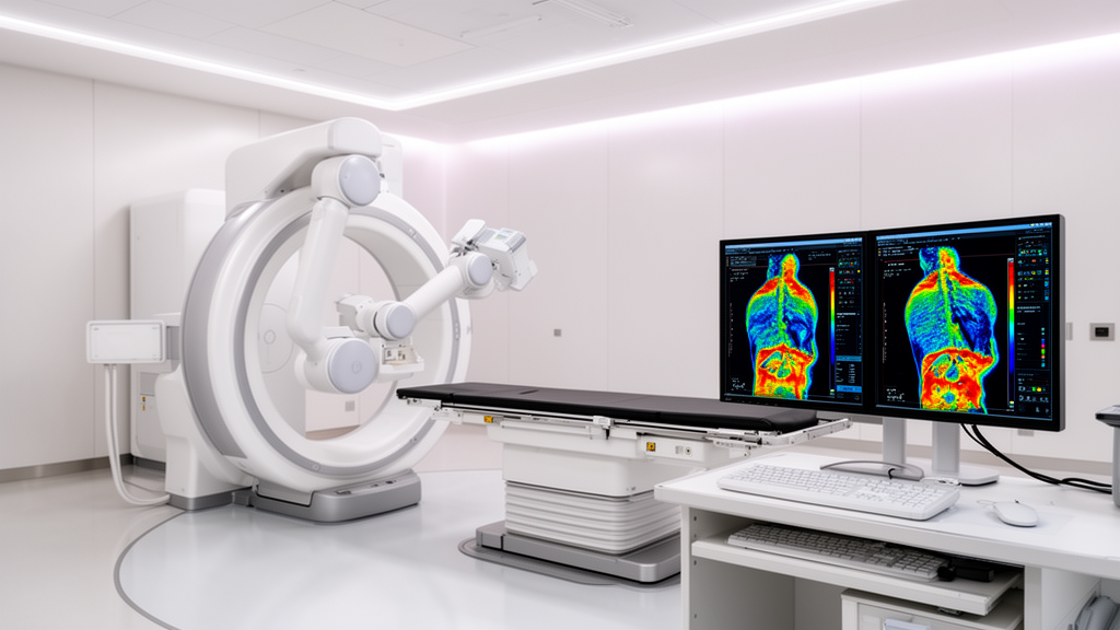
A retrospective investigation has demonstrated that machine learning classifiers trained on organ-specific dosiomic features can accurately predict late genitourinary adverse events in patients undergoing stereotactic body radiation therapy for localized prostate cancer. Published in BMC Cancer, the study evaluated treatment plans generated for CyberKnife robotic radiosurgery to determine whether advanced spatial radiation metrics could identify patients at risk of chronic urinary toxicity more effectively than conventional planning tools and clinical assessments.
The study evaluated a cohort of eighty-seven patients diagnosed with localized prostate adenocarcinoma who underwent extreme hypofractionation delivered via the CyberKnife system. Treatment regimens delivered total radiation doses ranging from 35 to 36.25 Gray administered across five treatment fractions. Following therapy, patients were monitored over an extended observation timeline spanning up to five years to document post-treatment complications.
The clinical endpoint examined by the investigators was the development of late genitourinary adverse events classified as grade 1 or higher according to the Radiation Therapy Oncology Group grading criteria. To ensure the analysis strictly addressed delayed complications rather than acute reactions, the primary endpoint was restricted to toxicities emerging beyond six months post-radiation. Within the analyzed patient group, late genitourinary adverse events were documented in 17.2 percent of individuals.
To capture fine-grained radiation distribution data, researchers employed the PyRadiomics package to extract high-dimensional dosiomic features from multiple anatomical contours. These regions of interest included the prostate gland, the planning target volume, the urinary bladder, the rectum, the penile bulb, and a surrogate urethra model. The investigators established four distinct predictive models built on identical patient datasets and partitions: one relying on clinical and patient-reported variables, one utilizing traditional dose-volume histogram metrics, one utilizing extracted dosiomics, and a fourth combining all parameter blocks.
Model training incorporated an eighty-twenty patient-level train-test partition combined with feature filtering and least absolute shrinkage and selection operator regression. Classifiers were trained applying group-aware cross-validation and class weighting to address class imbalance. Final model validation on the independent hold-out test set demonstrated that the dosiomic model achieved the strongest discriminative capability, recording a receiver operating characteristic area under the curve of 0.92 and a precision-recall area under the curve of 0.84 against a baseline prevalence reference of 0.167. In comparison, the combined model achieved an area under the curve of 0.90, the dose-volume histogram model achieved 0.86, and the clinical model achieved 0.46. Balanced accuracy reached 0.80 for dosiomics, outperforming the dose-volume metric score of 0.70 and the clinical model score of 0.43.
Further analysis revealed that integrating conventional dose-volume statistics and clinical variables into the dosiomic framework failed to improve discriminative accuracy over dosiomic parameters alone. To ensure clinical transparency, the investigators performed SHapley Additive exPlanations analysis, which isolated a compact set of interpretable risk predictors. These influential features captured both organ geometry and spatial dose heterogeneity across the bladder, urethra, and prostate. While these findings highlight the predictive value of dosiomic analysis in radiosurgery planning, the authors noted that multicenter validation remains necessary to establish model generalizability.
The evaluation incorporated bootstrap confidence intervals across the independent testing phase. For precision-recall metrics, the dosiomic model achieved a 95 percent confidence interval of 0.53 to 1.00, while the combined framework yielded an interval of 0.37 to 1.00. The dose-volume histogram model generated a confidence interval spanning 0.35 to 1.00, and the clinical model recorded a range between 0.16 and 0.37.
Regarding discrimination across receiver operating curves, the dosiomic approach demonstrated a 95 percent confidence interval spanning 0.69 to 1.00. Corresponding confidence intervals reached 0.73 to 1.00 for the combined strategy, 0.68 to 1.00 for dose-volume histograms, and 0.22 to 0.70 for clinical variables. The methodology also assessed feature selection stability across independent training sets and related SHAP contributions directly to conventional dose-volume metrics.
Subject of Research: Dosiomics-based machine learning prediction of late genitourinary toxicity following CyberKnife prostate SBRT
Article Title: Dosiomics-based prediction of late genitourinary side-effects after CyberKnife for prostate cancer: a SHAP-explainable machine learning approach
Article References: Mottareale, R., Serra, M., Pezzulla, D., Cilla, S., Borzillo, V., Scipilliti, E., Franzese, D., Perdonà, S., Passaro, F., Izzo, A., Rossetti, S., Pignata, S., Ametrano, G., d’Alesio, V., Buonanno, F., Arrichiello, C., Mercogliano, S., Di Franco, R., & Ravo, V. (2026). Dosiomics-based prediction of late genitourinary side-effects after CyberKnife for prostate cancer: a SHAP-explainable machine learning approach. BMC Cancer. https://doi.org/10.1186/s12885-026-17279-2
Image Credits: AI Generated
DOI: 10.1186/s12885-026-17279-2
Keywords: prostate cancer, CyberKnife, stereotactic body radiation therapy, dosiomics, machine learning, genitourinary toxicity, SHAP analysis, PyRadiomics, radiation oncology, dose-volume histogram, RTOG scale, prostate adenocarcinoma, treatment planning, predictive modeling, surrogate urethra model, spatial heterogeneity
News Source: Nathaniel Bowman. (November 17, 2026). Dosiomics Machine Learning Model Predicts Late Urinary Toxicity After Prostate CyberKnife SBRT. Scienmag.