


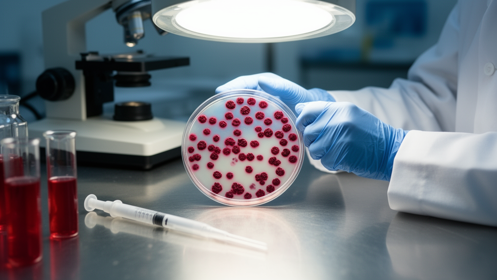
Researchers at Washington University in St. Louis have shown that a decades-old zinc-binding dye can solve one of the most stubborn problems in islet transplantation research: finding the graft after it has been placed in the body. In a study published in BMC Methods, James Lu, Matthew Ishahak, Marlie M. Maestas and Jeffrey R. Millman demonstrated that dithizone staining reliably marks insulin-producing cells transplanted under the skin of mice, turning otherwise invisible grafts into bright red targets that can be located by eye, excised and analyzed. The work pairs this staining method with an automated islet-counting system, creating a reproducible framework for dosing and recovering transplanted islets in preclinical experiments.
Pancreatic islet transplantation is a promising strategy for restoring insulin independence in patients with type 1 diabetes. For more than two decades, the standard approach has been to infuse primary human islets, isolated from the pancreases of deceased donors, through the hepatic portal vein. This route delivers the islets to the liver, where they can access the systemic circulation and mimic the way endogenous insulin reaches the organ. More recently, islets derived from human pluripotent stem cells, known as SC-islets, have shown promise in preclinical animal models and in a clinical trial. Yet both approaches face persistent obstacles, including poor long-term graft survival, blood-mediated inflammatory responses, immune-mediated destruction, and difficulty monitoring graft viability and function over time.
The invasive nature of portal vein transplantation also complicates the retrieval of islets and their longitudinal assessment, which hinders mechanistic studies aimed at improving graft survival. To sidestep these problems, preclinical researchers have explored alternative transplantation sites in mice, including the renal subcapsular space, the intramuscular region and the subcutaneous cavity. The kidney capsule is highly vascularized and easy to access in small animals, but it has limited translational capacity and is not a suitable location for human transplantation. The intramuscular space allows better vascular integration but may involve complex surgical implantation and fibrotic encapsulation. The subcutaneous space stands out for its minimally invasive access, potential for real-time monitoring, non-terminal graft retrieval and compatibility with devices. Its weakness is that it is relatively poorly vascularized, which challenges islet engraftment and survival.
Even when islets do engraft under the skin, locating them is remarkably difficult. The grafts disperse within the tissue and offer little visual contrast against the surrounding tissue, making retrieval impractical. This limitation has constrained the experimental utility of the subcutaneous site, despite its many practical advantages. The Millman laboratory’s answer exploits a property of insulin-producing beta cells: they contain unusually high concentrations of intracellular zinc. Dithizone, a zinc-chelating dye long used in vitro to assess islet purity and viability, binds selectively to that zinc and produces a distinctive red color. The team hypothesized that this same chemistry could serve as a rapid and cost-effective way to localize transplanted islets within the subcutaneous space.
The study’s second pillar addresses a quieter but equally important problem: measuring how many islets are actually transplanted. Standardization of transplanted islet volume, expressed as islet equivalents or IEQs, is essential for interpreting graft survival, functional outcomes and dosage responses across experimental groups. Manual IEQ quantification is labor-intensive and impractical when processing large numbers of samples, and these limitations introduce variability that reduces experimental reproducibility. The BioRep Islet Cell Counter, an automated image-based system, offers a standardized alternative by objectively measuring islet size distribution and total islet volume. Despite its growing adoption in islet research, its integration with transplantation and dosage-response relationships has remained underreported, particularly for subcutaneous models.
To build the framework, the researchers first generated SC-islets from the HUES8 stem cell line using a multistep differentiation protocol that recapitulates key stages of embryonic pancreatic development with temporally controlled growth factors and small molecules. They validated the differentiation by documenting islet morphology at the start of each key stage and confirmed function with a glucose-stimulated insulin secretion assay. Both primary human islets, which secreted 4.144 ± 0.055 µIU per 1,000 cells, and SC-islets, which secreted 1.086 ± 0.204 µIU per 1,000 cells, released insulin in response to high glucose. Consistent with previous reports, the SC-islets were functionally immature compared with primary islets, but the team confirmed they were within the range of expected secretion levels.
Before transplantation, islet volume was standardized using the BioRep counter. Aliquots of islets were transferred to a counting dish with sufficient medium or phosphate-buffered saline to keep clusters suspended, the dish was swirled to distribute the islets while minimizing overlap, since the system identifies overlapping clusters as a single islet, and the unstained profile was selected with red pixel calibration manually overridden. Non-islet debris was excluded before analysis, and the software generated a report detailing total IEQs and islet size distribution. The resulting doses were closely matched: mice receiving primary human islets got an average of 4,200 ± 227.7 IEQ, while those receiving SC-islets got 4,128 ± 158.7 IEQ. Approximately 4,000 IEQ of either cell type was injected into the subcutaneous space on the dorsal medial left side of male immunodeficient NOD.Cg-PrkdcscidIl2rgtm1Wjl/SzJ mice, five animals per group, using a 27-gauge needle.
Graft function was assessed longitudinally through weekly weight measurements and human C-peptide assays using an ultrasensitive ELISA. The islets maintained function in vivo, as demonstrated by detectable human C-peptide levels. At four weeks post-transplantation, primary human islets produced 170.208 ± 142.986 pmol/L of C-peptide compared with 59.247 ± 15.433 pmol/L for SC-islets, a difference that was not statistically significant. Body weights showed no significant differences following transplantation, except that at four weeks mice receiving SC-islets were significantly lighter than non-transplanted mice, with a p-value of 0.004. Because the mice were normoglycemic and immunodeficient rather than diabetic, metabolic rescue was not an experimental endpoint; the primary objective was to establish and validate the methodological framework for standardized dosing and reliable graft localization.
The staining procedure itself is strikingly simple. Four weeks after transplantation, the mice were euthanized and the skin containing the graft site was harvested. Samples were submerged in a 1 mg/mL dithizone solution, prepared by dissolving the dye in dimethyl sulfoxide, diluting it in phosphate-buffered saline and sterile-filtering it, and incubated for five minutes at room temperature. Before staining, neither primary human islet nor SC-islet grafts were readily identifiable to the naked eye. After soaking, the grafts became visible as red-stained regions marking the zinc-rich beta cells, and the team photographed them with an iPhone 15 Pro Max before excising the tissue. Immunohistochemical validation followed: sections stained for DAPI, C-peptide, chromogranin A, glucagon and somatostatin showed clear and specific labeling for all markers, with no apparent loss of signal or alteration in cellular distribution, confirming that dithizone does not interfere with downstream immunostaining or antigen detection.
The authors are careful about the method’s boundaries. Dithizone-based localization is inherently limited to terminal analyses and cannot be applied to living animals, so it complements rather than replaces non-invasive imaging; longitudinal monitoring can be achieved in preclinical designs by periodically sacrificing subsets of animals at defined time points. The approach has been validated only for short-term exposure in terminally collected samples, since prolonged dithizone incubation has been reported to adversely affect islet viability, and compatibility with molecular assays such as RNA sequencing or proteomics was not evaluated. Future studies could assess the effects of dithizone exposure, test diabetic models under hyperglycemic stress, and explore other transplantation sites and imaging modalities. Within those limits, the method gives researchers a rapid, cost-effective tool for recovering IEQ-normalized grafts at defined endpoints, supporting more systematic comparison of islet dosage, graft composition and experimental conditions as stem cell-derived islets advance toward scalable therapeutic products.
Subject of Research: A dithizone staining method combined with automated islet equivalent quantification for detecting and retrieving subcutaneously transplanted islet grafts in mice
Article Title: Optimizing post-transplantation detection of subcutaneously transplanted islets using dithizone staining
Article References: Lu, J., Ishahak, M., Maestas, M. M., & Millman, J. R. (2026). Optimizing post-transplantation detection of subcutaneously transplanted islets using dithizone staining. BMC Methods, 3(1), Article 23. https://doi.org/10.1186/s44330-026-00075-6
Image Credits: AI Generated
DOI: 10.1186/s44330-026-00075-6
Keywords: islet transplantation, dithizone staining, type 1 diabetes, stem cell-derived islets, subcutaneous graft localization, islet equivalents, BioRep Islet Cell Counter, beta cells, immunohistochemistry, C-peptide, preclinical models, graft recovery
News Source: Drew Townsend. (October 30, 2026). Dithizone Dye Makes Transplanted Islets Visible for Retrieval. Scienmag.



