
A groundbreaking study published in the Journal of Thoracic Disease heralds a transformative approach in pulmonary segmentectomy, introducing inter-multisegmental veins (IMSVs) as a pivotal anatomical landmark for surgical decision-making. This innovative research challenges the traditional reliance solely on radiological lesion characteristics and spotlights the spatial relationship between lung lesions and IMSVs, ushering in a paradigm shift in the management of early-stage non-small cell lung cancer (NSCLC).
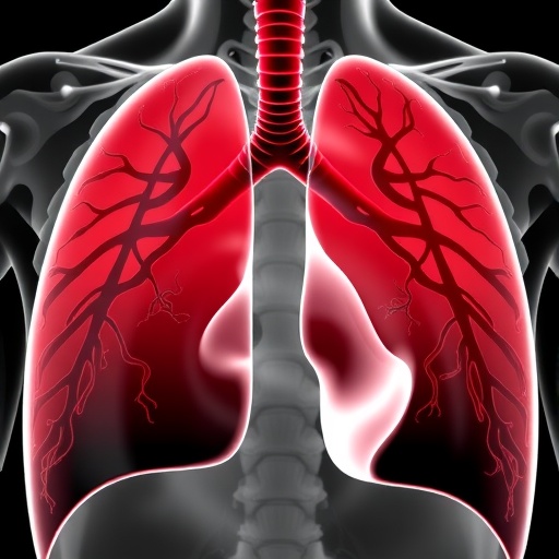
The anatomy of pulmonary veins has long been recognized as complex yet critically important in thoracic surgery. IMSVs, blood vessels running between lung segments, have previously been underappreciated in surgical planning. The study highlights the high prevalence of IMSVs and elaborates on their diverse drainage patterns, particularly of the lateral vein (Vl), contrasting sharply with the static nature of branches stemming from the superior segmental vein (V6b). Such vascular variations, once overlooked, play a subtle but essential role in determining optimal resection boundaries.
Central to the study is the identification of two essential positional indicators that refine sublobar resection candidacy: the depth ratio of lesions within the lung parenchyma and the lesion’s relative location to IMSVs along the transverse plane. The depth ratio quantifies how deeply a tumor resides from the visceral pleura towards the bronchial tree, providing a nuanced view of its accessibility via segmentectomy. Meanwhile, the lesion’s proximity to IMSVs offers a novel transverse positional parameter, allowing surgeons to anticipate and preserve vital venous structures, thereby enhancing surgical precision.
Historically, surgical decisions for sublobar resection in early-stage NSCLC hinged predominantly on traditional radiographic markers such as lesion size and consolidation-to-tumor ratio. These metrics, while valuable, offer limited spatial context and often neglect the three-dimensional venous anatomy that critically underpins segmental boundaries. Incorporating IMSVs into the surgical algorithm paves the way for a more anatomically attuned, patient-specific approach, potentially reducing unexpected intraoperative complications and achieving clear resection margins with greater consistency.
The implications of these findings extend beyond anatomical curiosity. Ensuring that lesions are aligned with IMSVs spatially facilitates effective anatomical segmentectomy — a lung-sparing surgery with outcomes comparable to lobectomy for appropriate tumor stages. The integration of IMSV mapping into preoperative planning enhances the surgeon’s ability to maintain oncological rigor while minimizing loss of healthy lung tissue, thus improving postoperative pulmonary function and quality of life.
This study’s observational design involved meticulous preoperative imaging and intraoperative validation, where the researchers correlated three-dimensional computed tomography reconstructions with real-time surgical anatomy. Such comprehensive methodology underscores the translational potential of vascular mapping, transforming theoretical knowledge into practical guidelines. Moreover, the variability observed in IMSV anatomy, especially in the lateral vein’s diverse drainage routes, suggests that individualized surgical planning must transcend generic templates and embrace patient-specific venous architecture.
The recognition of IMSVs as indispensable positional indicators tackles a critical challenge: defining safe surgical margins in a minimally invasive yet oncologically sound manner. By accounting for both longitudinal depth and transverse location, surgeons can make more informed intraoperative decisions, minimizing margin-positive resections that are associated with higher local recurrence rates. This precision is paramount in clinical T1a–bN0 NSCLC patients, where sublobar resection is increasingly viewed as a favorable alternative to traditional lobectomy.
Furthermore, this anatomical refinement harmonizes with advances in thoracic surgical technology, including video-assisted thoracoscopic surgery (VATS) and robotic-assisted techniques, which demand precise navigation of segmental planes. The integration of IMSV positional data into surgical planning software could soon aid surgeons in constructing real-time vascular maps, enhancing both the safety and efficiency of segmentectomy procedures.
The study also brings attention to a previously underreported vascular element—the lateral vein (Vl)—whose variability represents a biomechanical and surgical challenge. Unlike the relatively constant superior segmental vein branches, the lateral veins’ heterogeneity necessitates a careful, tailored approach to preserve venous drainage and prevent postoperative complications such as segmental congestion or infarction. This nuanced understanding exemplifies the importance of personalized surgery in the current era.
Beyond its technical merits, the research advocates for a shift in surgical education and training. Surgeons must familiarize themselves with the topographical subtleties of IMSVs and incorporate three-dimensional preoperative imaging assessment into routine practice. This evolution will foster improved interdisciplinary collaboration between radiologists, thoracic surgeons, and oncologists, ultimately benefiting patient outcomes and resource utilization.
In conclusion, the pioneering work on IMSVs as new positional indicators for pulmonary segmentectomy represents a significant advance in thoracic oncology surgery. By refining the concepts of longitudinal and transverse tumor localization relative to critical venous structures, the study offers a novel framework for safer and more effective sublobar lung resections. As the prevalence of early-stage NSCLC diagnoses rises due to improved screening protocols, such anatomical insights are poised to transform surgical standards and elevate patient care to unprecedented levels.
Subject of Research: People
Article Title: Inter-multisegmental veins (IMSVs): a new positional indication for pulmonary segmentectomy
News Publication Date: 21-Feb-2025
Web References: http://dx.doi.org/10.21037/jtd-24-1799
References: Bian C, Fu C, Wang Y, Huang J, Yuan M, Chen L, Wang Q, Wang J. Inter-multisegmental veins (IMSVs): a new positional indication for pulmonary segmentectomy. J Thorac Dis 2025;17(2):603-613. doi: 10.21037/jtd-24-1799
Keywords: Surgery, Pulmonary Segmentectomy, Inter-multisegmental veins, Lung cancer, Non-small cell lung cancer (NSCLC), Anatomical resection, Thoracic surgery
Tags: anatomical landmarks in thoracic surgeryIMSVs in pulmonary surgeryinnovative surgical techniquesinter-multisegmental veinslung lesion depth rationon-small cell lung cancer managementprecision pulmonary segmentectomypulmonary vein drainage patternssublobar resection indicatorssurgical planning for lung cancerthoracic disease researchvascular anatomy in lung resection

