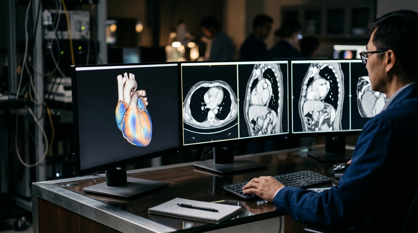
Accurate measurement of blood flow to the heart muscle often begins with a step that is easy to overlook: drawing the boundaries of the heart itself. In single-photon emission computed tomography myocardial perfusion imaging, known as SPECT MPI, quantitative analysis relies on polar maps that divide the left ventricle into segments and assign each one an uptake value. Those maps are only as good as the myocardial contours beneath them, and in commercial software those contours frequently demand significant manual adjustment, especially when a patient has severe perfusion defects or bright uptake from organs outside the heart.
A research team led by Hussein Akafzade and Jaewon Yang, publishing in EJNMMI Physics, reports a deep learning approach designed to remove that manual burden. Their model performs myocardial segmentation using non-contrast-enhanced CT images, the attenuation correction scans that are already acquired alongside SPECT MPI studies but that normally contain little contrast between the heart muscle and neighboring structures. The work is described as original research and was published as an open access article on 3 November 2026.
The methodological route the investigators took is notable for its economy. Rather than laboriously hand-drawing myocardial masks on CT images, they generated training labels by simply thresholding PET images with uniform myocardial uptake from 125 18F-FDG PET/CT studies. Because fluorodeoxyglucose accumulates relatively evenly in healthy heart muscle in these studies, the PET signal itself served as a template for where the myocardium lies, and the corresponding non-contrast CT images became the network’s input. The team configured an optimal network through the nn-UNet pipeline, a self-configuring framework that automatically tunes architecture, preprocessing and training settings to a given segmentation task.
Evaluation proceeded in stages. The network was first tested on 50 99mTc-sestamibi SPECT/CT MPI studies with uniform myocardial uptake, a different tracer and a different scanner modality from the training data, which tests whether the learned segmentation generalizes across imaging protocols. Performance was quantified with the Dice Similarity Coefficient, a standard overlap metric that ranges from 0, indicating no agreement, to 1, indicating perfect agreement between two segmentations.
The numbers the team reports are consistent across both validation and test data. DL-masks achieved a Dice Similarity Coefficient of 0.866 ± 0.035 in the PET/CT validation set and 0.846 ± 0.026 in the SPECT/CT test set. An overlap above 0.84 on out-of-modality test data indicates that the network captured the myocardial volume with a high degree of spatial agreement, despite the fact that the CT images it saw at test time came from SPECT/CT rather than PET/CT acquisitions.
The more clinically pointed test involved a harder population. The investigators selected an additional 50 SPECT MPI studies with severe myocardial defects or high extracardiac uptake, precisely the cases in which conventional software struggles and manual correction consumes the most time. For these studies, the researchers compared segmental uptake values in polar maps computed with and without the deep learning masks. The comparison revealed meaningful quantitative differences of approximately 10% in basal segments near the septum, compared with mid-cavity or apical segments. That regional pattern matters because the basal septum sits adjacent to the valve plane, an anatomical landmark that automated and manual contouring approaches frequently place differently, and errors there propagate directly into the segmental values that cardiologists read.
To assess the practical effect on workflow, three nuclear cardiologists visually compared myocardium contours with and without DL-masks in commercial software. Their Likert scoring demonstrated that the deep learning masks improved septal and valve plane placement, resulting in accurate myocardial segmentation without manual adjustment. In other words, the model did not merely reproduce a contour; it corrected the specific anatomical placement errors that force technologists and physicians to intervene by hand.
The clinical significance lies in what the result removes from the pipeline. Quantitative SPECT MPI is increasingly used to track perfusion numerically, compare studies over time and support decisions about revascularization, but its reliability depends on reproducible segmentation. When severe defects shrink the visible myocardial signal or extracardiac activity from the liver, bowel or other structures bleeds into the image, automated contouring tools can misplace the septal border or the valve plane, and every downstream segmental value shifts. A segmentation model that runs on the non-contrast CT already present in the acquisition, without requiring contrast injection or manual editing, addresses that failure mode at its source.
The study’s design also carries limitations worth noting. The training labels were derived from PET studies with uniform myocardial uptake, so the network learned from anatomically normal-appearing myocardium even though it was evaluated on studies with defects. The reported Dice coefficients, while strong, describe overlap with thresholded PET masks rather than an independent gold standard, and the visual comparison involved three cardiologists at a single institution. The authors nevertheless conclude that they demonstrated a DL segmentation model for accurate myocardial segmentation without manual adjustment and corresponding polar map-based quantification, particularly in studies with severe myocardial defects or high extracardiac uptake, the patient population where quantitative cardiac SPECT has historically been most fragile.
Subject of Research: Deep learning-based myocardial segmentation from non-contrast CT for quantitative SPECT myocardial perfusion imaging
Article Title: Deep learning-based myocardial segmentation using non-contrast-enhanced CT for quantitative SPECT myocardial perfusion imaging
Article References: Akafzade, H., Agusala, K., Vigen, R., Chandra, A., Chon, K. S., Rashidykanan, H., Seo, Y., Park, M.-A., & Yang, J. (2026). Deep learning-based myocardial segmentation using non-contrast-enhanced CT for quantitative SPECT myocardial perfusion imaging. EJNMMI Physics. https://doi.org/10.1186/s40658-026-00962-5
Image Credits: AI Generated
DOI: 10.1186/s40658-026-00962-5
Keywords: deep learning, myocardial segmentation, SPECT, myocardial perfusion imaging, non-contrast CT, polar map quantification, PET/CT, nn-UNet, nuclear cardiology, 99mTc-sestamibi, Dice similarity coefficient, extracardiac uptake
News Source: Blake Davidson. (November 5, 2026). Deep learning segments heart muscle on non-contrast CT for SPECT perfusion quantification. Scienmag.