In a remarkable leap forward for oncology and medical imaging, a team of researchers has developed a deep learning (DL) model that harnesses ultrasound imaging to predict platinum resistance in patients afflicted with epithelial ovarian cancer (EOC). This cutting-edge innovation is poised to transform treatment paradigms by enabling clinicians to anticipate therapeutic resistance, thereby tailoring interventions more effectively and improving patient outcomes in this aggressive malignancy.
Epithelial ovarian cancer remains one of the deadliest gynecological cancers worldwide, often diagnosed at an advanced stage and commonly treated with platinum-based chemotherapies. Unfortunately, a significant subset of patients develops resistance to platinum drugs, a phenomenon that severely compromises treatment efficacy and survival rates. Conventional approaches to foresee platinum resistance have largely been invasive or reliant on molecular profiling, which may not always be feasible in routine clinical practice.
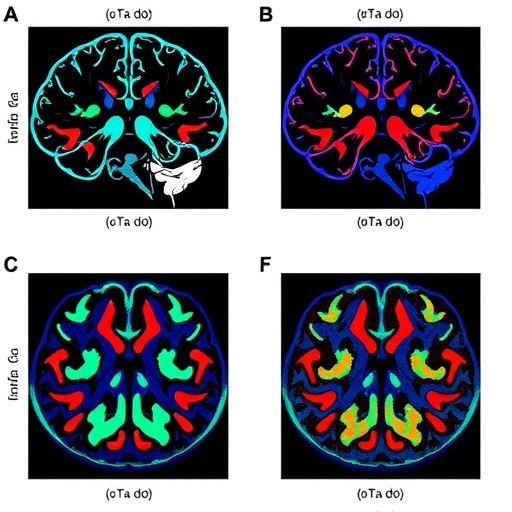
The research, conducted through a retrospective analysis, leveraged data from 392 patients diagnosed with EOC from 2014 to 2020. Prior to initial treatment, all subjects underwent pelvic ultrasound scanning, thus providing a rich repository of imaging data. The investigators ingeniously applied deep learning algorithms to analyze these ultrasound images, aiming to discern subtle patterns and features imperceptible to the human eye but indicative of the tumor’s chemoresistance profile.
Deep learning, a subdivision of artificial intelligence mimicking neuronal networks, has proven extraordinarily powerful in image recognition tasks. In this context, the researchers trained their DL model on the imaging data, enabling it to classify tumors likely to exhibit platinum resistance. The training involved feeding the model with input-output pairs: ultrasound images labeled as platinum-sensitive or platinum-resistant based on clinical follow-up. Through backpropagation and iterative optimization, the model refined its predictive capacity.
The model’s performance was rigorously assessed, employing receiver operating characteristic (ROC) curves to quantify diagnostic accuracy. Impressively, the area under the curve (AUC) reached 0.86 in both internal and external test sets, underscoring the model’s robustness and reproducibility across different patient cohorts. An AUC of 0.86 signifies a high level of discriminative ability, confirming that the DL system can effectively differentiate between resistant and sensitive tumors based solely on ultrasound imaging.
To ensure the model’s clinical utility transcended statistical validation, decision curve analysis (DCA) was performed. This technique evaluates the net benefit of a diagnostic tool across varying threshold probabilities, revealing that the DL model offers significant clinical value in guiding treatment decisions. Furthermore, calibration curves confirmed the model’s predictive outputs were well-aligned with actual patient outcomes, an essential criterion for trustworthiness in clinical settings.
Beyond its predictive prowess, the DL model demonstrated prognostic significance. Kaplan–Meier survival analyses highlighted that patients classified into the high-risk group for platinum resistance experienced significantly worse recurrence-free survival. Hazard ratios of approximately 3.0 in both internal and external validation cohorts confirmed that the model’s optimal cutoff reliably identifies patients with a markedly elevated risk of early relapse, enabling oncologists to stratify patients more precisely.
This novel approach offers profound implications for personalized medicine. By integrating non-invasive ultrasound imaging with advanced artificial intelligence, physicians could foresee platinum resistance before commencing chemotherapy. Such foresight would empower clinicians to modify treatment regimens proactively, potentially incorporating alternative chemotherapeutic agents, targeted therapies, or novel clinical trial enrollment, thereby maximizing therapeutic efficacy and sparing patients from unnecessary side effects.
The study meticulously adhered to rigorous methodological standards, utilizing an extensive and well-characterized patient cohort, which strengthens the generalizability of findings. Additionally, the inclusion of both internal and external validation sets mitigates overfitting concerns, a common challenge in AI model development. These methodological strengths propel the model closer to eventual clinical deployment.
While the research presents compelling evidence, several considerations warrant further exploration. Ultrasound image quality can vary based on operator skill and equipment, potentially affecting model input consistency. Future studies might explore standardization protocols or augmented imaging techniques to enhance model reliability. Moreover, integrating multi-modality data, such as genomic or serological markers, with ultrasound-based DL predictions could further refine resistance forecasting.
The convergence of deep learning with accessible imaging modalities like ultrasound signals a paradigm shift in oncology diagnostics. Unlike magnetic resonance or computed tomography scans, ultrasound is widely available, cost-effective, and free of ionizing radiation, making it an ideal candidate for broad clinical implementation. This democratization of advanced diagnostic tools could dramatically impact patient care, especially in resource-limited settings.
Moreover, transparent reporting of model interpretability remains vital. While deep learning models achieve high accuracy, the “black box” nature often obscures reasoning pathways. Integration of explainable AI techniques to elucidate imaging features driving predictions would enhance clinician trust and facilitate regulatory approval.
In sum, this pioneering work illustrates the transformative potential of artificial intelligence applied to routine ultrasound imaging for anticipating chemotherapy resistance in epithelial ovarian cancer. By bridging technological innovation with clinical necessity, the study charts a promising course toward tailored oncologic therapies that improve outcomes and optimize healthcare resources.
As the oncology community eagerly anticipates further validation and prospective trials, this development heralds a new era where machine learning models augment clinical acumen, advancing personalized medicine from concept to reality. Integrating such AI-driven tools into standard care pathways could redefine how epithelial ovarian cancer is managed, shifting the focus from reactive treatment to proactive, precision-guided intervention.
Subject of Research: Prediction of platinum resistance in epithelial ovarian cancer using deep learning applied to ultrasound imaging.
Article Title: Deep learning based on ultrasound images to predict platinum resistance in patients with epithelial ovarian cancer.
Article References:
Su, C., Miao, K., Zhang, L. et al. Deep learning based on ultrasound images to predict platinum resistance in patients with epithelial ovarian cancer. BioMed Eng OnLine 24, 58 (2025). https://doi.org/10.1186/s12938-025-01391-8
Image Credits: AI Generated
DOI: https://doi.org/10.1186/s12938-025-01391-8
Tags: Deep Learning in Oncologyepithelial ovarian cancer treatment advancementsimproving patient outcomes in EOCinnovative imaging techniques in oncologymachine learning applications in medicinenon-invasive methods in cancer diagnosisovercoming chemotherapy resistancepredicting platinum resistance in ovarian cancerretrospective analysis of ovarian cancer datatailored interventions for cancer patientstransforming cancer treatment paradigmsultrasound imaging for cancer prediction

