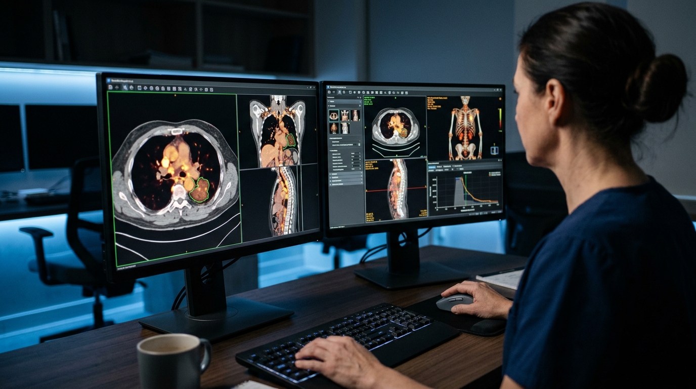
A disease-specific deep learning model designated HL4-Net accurately automates the determination of metabolic tumor volume in Hodgkin lymphoma, achieving segmentation and predictive performance equivalent to manual measurements. While metabolic tumor volume derived from positron-emission tomography imaging has demonstrated predictive value, its clinical application remains limited, leading researchers to develop and validate a machine learning model for fully automated segmentation in Hodgkin lymphoma.
The investigation utilized a total of 1,558 positron-emission tomography scans, including 544 baseline scans with manually segmented tumor volumes from multicenter German Hodgkin Study Group HD16, HD17, and HD18 trials. A residual encoder U-Net deep learning architecture was trained on lymphoma-specific trial data to create HL-Net. For comparison, models were trained on a multicancer dataset of 1,014 scans separately as Auto-PET, and in combination with lymphoma scans as HL/Auto-PET. An additional lymphoma-specific variant, HL4-Net, incorporated an auxiliary input mask set at a threshold of SUV 4 or greater. All models were evaluated using a hold-out cohort from non-overlapping external trial sites.
In validation testing, the lymphoma-specific HL-Net achieved a higher Dice score, sensitivity, and positive predictive value compared to the multicancer Auto-PET model, recording a Dice score of 0.865 versus 0.661 (p<0.001), sensitivity of 1.000 versus 0.727 (p<0.001), and positive predictive value of 1.000 versus 0.600 (p<0.001). The HL4-Net architecture further improved segmentation performance, reaching a Dice score of 0.949 (p<0.001). Machine-learning-derived metabolic tumor volumes from HL4-Net demonstrated a very strong correlation with manual measurements, with an R-squared value of 0.981 (p<0.001).
When assessing clinical utility in predicting early treatment response, metabolic tumor volumes determined by HL4-Net achieved an area under the curve of 0.791, demonstrating comparable performance to manual segmentation, which achieved an area under the curve of 0.799 (p=0.649). The authors concluded that HL4-Net was optimal for high-accuracy lesion segmentation, highlighting the necessity of disease-specific architectures and establishing that automated volumes can serve as an effective surrogate for manual measurements.
Subject of Research: Automated metabolic tumor volume segmentation in Hodgkin lymphoma
Article Title: Fully automated determination of metabolic tumor volume in Hodgkin lymphoma
Article References: Ferdinandus, J., Clement, C., Tharmaseelan, H., Koller, P., Lopes, L., Novak, U., Pabst, T., Lommen, M., Pandya, K., Roth, K., Rominger, A., Shi, K., Borchmann, P., Kobe, C., & Seifert, R. (2026). Fully automated determination of metabolic tumor volume in Hodgkin lymphoma. EJNMMI Physics. https://doi.org/10.1186/s40658-026-00935-8
Image Credits: AI Generated
DOI: 10.1186/s40658-026-00935-8
Keywords: Hodgkin lymphoma, metabolic tumor volume, positron emission tomography, machine learning, deep learning, image segmentation, residual encoder U-Net, treatment response prediction, Fully, automated, determination, metabolic
News Source: Nathaniel Bowman. (November 5, 2026). Deep Learning Model Automates Metabolic Tumor Volume Segmentation in Hodgkin Lymphoma. Scienmag.