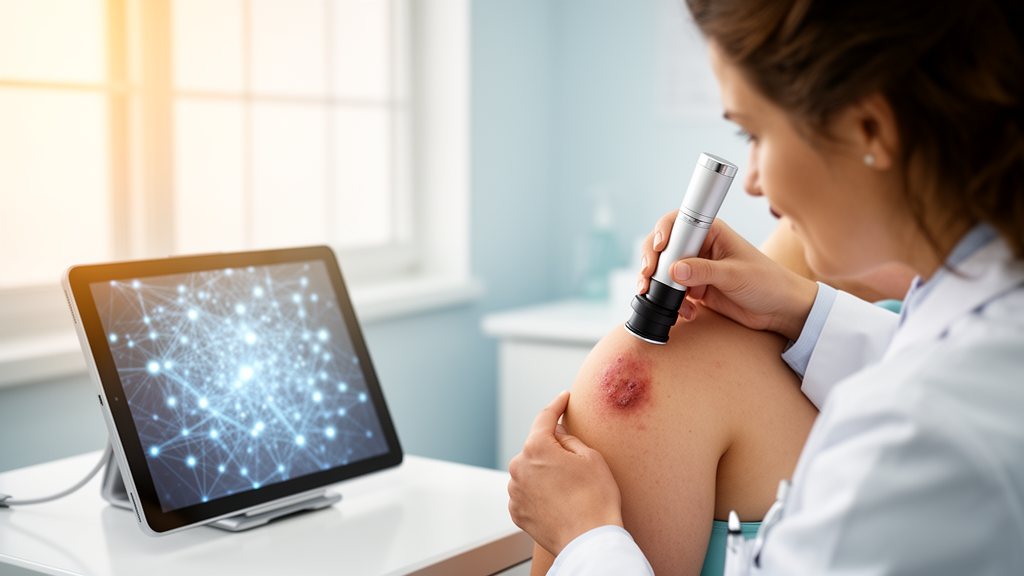
Artificial intelligence has become one of the most powerful tools in dermatology, capable of carving lesions out of dermoscopic images and sorting them into diagnostic categories in seconds. Yet a new structured narrative review published in Frontiers in Artificial Intelligence argues that the field’s headline accuracy numbers tell only a fraction of the story. The study, led by EBE Babu and SMS Murali of the School of Computer Science and Engineering at Vellore Institute of Technology in India, surveys journal papers published mainly from 2018 to 2026 and connects every stage of the pipeline, from raw image acquisition to clinical deployment, into a single critical analysis. Its central message is sobering: performance figures obtained on different datasets, data partitions and experimental setups cannot be directly compared, and a model that dazzles on a benchmark may fail quietly in a clinic.
The motivation is straightforward. Manual dermoscopic analysis is time-consuming, depends heavily on specialist expertise and is vulnerable to inter-observer variability, so reliable computer-aided diagnostic tools are badly needed. Early diagnosis of skin cancer is associated with more favorable treatment options and increased survival, which raises the stakes for automation. Convolutional neural networks have proven adept at picking out discriminating features in dermoscopic images, and architectures such as U-Net, ResNet, DenseNet and EfficientNet have repeatedly shown effectiveness in both pixel-level segmentation and diagnostic classification. But skin lesions vary enormously in size, shape, color, texture, pigmentation and borders, and artifacts such as hair, rulers, bubbles, shadows and inconsistent lighting can push models toward learning irrelevant image properties rather than genuine pathology.
A large share of the field’s progress rests on publicly available annotated databases. The HAM10000 dataset, with 10,015 multi-source dermoscopic images covering seven types of pigmented skin lesions, has become the workhorse for testing classification systems, with diagnoses validated through histopathology, expert consensus, follow-up or confocal microscopy. The smaller PH2 dataset, containing benign and atypical nevi and melanoma with expert annotations and manual delineations, is often reserved for external validation, while Derm7pt pairs dermoscopic and clinical images with structured seven-point checklist information, making it valuable for multimodal evaluation. The International Skin Imaging Collaboration archive, continuously extended and feeding numerous challenges, underpins much of the comparative work, although the review stresses that ISIC-2016, 2017, 2018 and 2019 differ in size, class distribution, annotation procedure and evaluation protocol, so results across them are not equivalent.
The review devotes detailed attention to preprocessing, warning that it should never be treated as a fixed recipe. Histogram equalization and CLAHE can improve contrast but may also amplify hair, bubbles, rulers and compression artifacts. Color normalization can harmonize images across devices and institutions yet risk suppressing diagnostically meaningful pigmentation patterns. Hair removal through morphological filtering or GAN-based inpainting, investigated by Bansal and Sridhar in 2024, must be applied cautiously because vessels, pigment networks and streaks can resemble hair, and generated texture may be artificial. The authors propose four practical pipelines tailored to standard transfer-learning classification, boundary-oriented segmentation, transformer-based analysis and cross-dataset or multi-institutional studies, insisting that every preprocessing choice be validated through downstream segmentation or classification performance rather than visual appeal.
On the segmentation side, the review traces an arc from thresholding, edge detection, region growing, clustering and active contours to fully convolutional networks, U-Net and its many descendants. Traditional methods remain computationally cheap and relatively interpretable but depend on manually selected parameters that may not transfer between datasets. U-Net’s encoder-decoder design with skip connections works well even with limited annotated data, and variants such as Attention U-Net, U-Net++ and MultiResUNet refine boundary localization, though added complexity demands ablation studies under identical conditions. More recently, transformer architectures have entered the arena: TransUNet merges convolutional feature extraction with global self-attention, while skin-lesion-specific models such as SkinFormer, which models statistical texture through kurtosis-guided counting, achieved 93.2 percent Dice on ISIC-2018, and Xin and colleagues’ transformer-guided self-adaptive multi-scale network targeted lesions of varying size.
Foundation models add a new dimension. The Segment Anything Model introduced promptable segmentation using points, boxes and masks, and MedSAM extended this approach to medical image-mask data with internal and external validation on a large collection that included dermoscopy. The review is careful about what this means: the remaining concern is not a lack of dermoscopic data in MedSAM’s development, but the need for independent external validation on dermoscopic datasets distinct from those used to build the foundation model. Promptable tools may support annotation, physician-assisted correction and semi-automatic segmentation, but natural-image pretraining can produce inadequate masks for low-contrast lesions, hair occlusion and uneven pigmentation, so these models should be seen as complementary instruments rather than automatic replacements for specialist networks.
Classification research shows a parallel evolution. Conventional machine learning with handcrafted color, shape and texture descriptors and classifiers such as support vector machines and random forests still suits small datasets and resource-limited settings. CNN backbones from VGG to MobileNet dominate modern work, with transfer learning reducing training demands, although natural-image pretraining introduces domain mismatch. Representative results illustrate both promise and caveats: a customized CNN reached 95.18 percent accuracy on HAM10000 with augmented data and k-fold cross-validation, ResNet-50 achieved 89.28 percent weighted accuracy on ISIC-2018 within a segmentation-guided pipeline, and a double-branch network hit 91.58 percent accuracy and 0.985 AUC on HAM10000 while still struggling with minority categories such as melanoma. Vision Transformers, ensembles and hybrid segmentation-classification systems such as S2C-DeLeNet round out the landscape, each carrying costs in data, computation and interpretability.
Perhaps the review’s most consequential contribution is its critique of evaluation practice. Overall accuracy can be badly misleading under class imbalance, since benign lesions dominate most datasets and a classifier can score highly while missing rare malignancies. The authors call for balanced accuracy, class-wise sensitivity and specificity, F1-scores, confusion matrices, appropriately averaged AUC, calibration measures such as expected calibration error and Brier score, confidence intervals and, crucially, external-dataset and subgroup performance. Patient-level separation is required wherever patient identifiers exist, so that related images do not appear in both training and testing, and external validation on an independent dataset is a far stronger signal of generalization than a random split of a single benchmark. Shortcut learning through rulers, markings and borders receives particular emphasis as a silent corruptor of benchmark results.
The challenges ahead are formidable. Class imbalance, dataset and device bias, domain shift, limited representation of darker skin tones, inadequate external validation, uncertain calibration, limited explainability of efficient models and the heavy computational demands of transformers and ensembles all stand between current benchmarks and trustworthy clinical decision making. Explainable AI methods such as Grad-CAM and SHAP, combined in frameworks like MultiExCam, can help assess whether predictions rest on lesion characteristics or artifacts, but visually plausible explanations do not prove faithful reasoning. Emerging directions include self-supervised learning, multimodal fusion of dermoscopic images with clinical metadata, federated learning, continual learning for post-deployment adaptation and lightweight architectures for mobile and point-of-care use, with the firm caveat that efficiency must never compromise sensitivity to malignant or rare lesions.
The review’s final verdict is measured rather than triumphal. Deep learning has genuinely advanced skin lesion segmentation and classification, and CNN and U-Net methods remain practical for many dermoscopic datasets while transformers and hybrids offer stronger global-context modeling at higher cost. But the leap from benchmark to bedside requires simultaneous attention to safety, robustness, fairness, interpretability, reproducibility, efficiency and demonstrated clinical utility, ideally through multi-center external and prospective validation and human-AI partnership studies that compare collaborative performance against human-only and model-only baselines. Automated systems, the authors conclude, should support rather than replace clinical expertise, and future progress should be measured not by isolated benchmark gains but by evidence that these tools improve diagnostic sensitivity, referral decisions, workload and, ultimately, patient outcomes.
Subject of Research: A structured narrative review of deep learning methods for skin lesion segmentation and classification, covering datasets, preprocessing, architectures, evaluation, fairness and clinical translation.
Article Title: A comprehensive study of skin lesion segmentation and classification using deep learning techniques
Article References: Babu, E., & Murali, S. (2026). A comprehensive study of skin lesion segmentation and classification using deep learning techniques. Frontiers in Artificial Intelligence, 9, Article 1907266. https://doi.org/10.3389/frai.2026.1907266
Image Credits: AI Generated
DOI: 10.3389/frai.2026.1907266
Keywords: deep learning, skin lesion, dermoscopy, segmentation, classification, convolutional neural networks, U-Net, Vision Transformers, explainable AI, external validation, class imbalance, foundation models
News Source: Blake Davidson. (October 29, 2026). Deep learning meets dermatology: new review maps the road from skin lesion AI benchmarks to bedside reality. Scienmag.