• HOME
  • NEWS
  • EXPLORE
    • CAREER
      • Companies
      • Jobs
    • EVENTS
    • iGEM
      • News
      • Team
    • PHOTOS
    • VIDEO
    • WIKI
  • BLOG
  • COMMUNITY
    • FACEBOOK
    • INSTAGRAM
    • TWITTER
Sunday, March 15, 2026
BIOENGINEER.ORG
No Result
View All Result
  • Login
  • HOME
  • NEWS
  • EXPLORE
    • CAREER
      • Companies
      • Jobs
        • Lecturer
        • PhD Studentship
        • Postdoc
        • Research Assistant
    • EVENTS
    • iGEM
      • News
      • Team
    • PHOTOS
    • VIDEO
    • WIKI
  • BLOG
  • COMMUNITY
    • FACEBOOK
    • INSTAGRAM
    • TWITTER
No Result
View All Result
Bioengineer.org
No Result
View All Result
Home NEWS Science News Health

Deep learning differentiates small renal masses on multiphase CT

Bioengineer by Bioengineer
January 10, 2020
in Health
Reading Time: 2 mins read
0
IMAGE
Share on FacebookShare on TwitterShare on LinkedinShare on RedditShare on Telegram

Deep learning method with neural network accurately differentiates between malignant and benign solid masses on contrast-enhanced CT scans, says AJR

IMAGE

Credit: American Journal of Roentgenology (AJR)

Leesburg, VA, January 10, 2020–A deep learning method with a convolutional neural network (CNN) can support the evaluation of small solid renal masses in dynamic CT images with acceptable diagnostic performance, according to an article published ahead-of-print in the March issue of the American Journal of Roentgenology (AJR).

Between 2012 and 2016, researchers at Japan’s Okayama University studied 1807 image sets from 168 pathologically diagnosed small (? 4 cm) solid renal masses with four CT phases–unenhanced, corticomedullary, nephrogenic, and excretory–in 159 patients.

Masses were classified as malignant (n = 136) or benign (n = 32) using a 5-point scale, and this dataset was then randomly divided into five subsets.

As lead AJR author Takashi Tanaka explained, “four were used for augmentation and supervised training (48,832 images), and one was used for testing (281 images).”

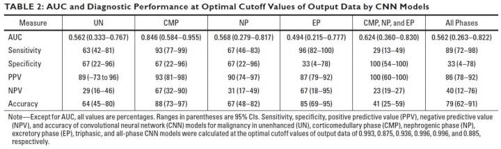
Utilizing the Inception-v3 architecture CNN model, the AUC for malignancy and accuracy at optimal cutoff values of output data were evaluated in six different CNN models.

Finding no significant size difference between malignant and benign lesions, Tanaka’s team did find that the AUC value of the corticomedullary phase was higher than that of other phases (corticomedullary vs excretory, p = 0.022).

Additionally, the highest accuracy (88%) was achieved in the corticomedullary phase images.

Multivariate analysis revealed that the CNN model of corticomedullary phase was a significant predictor for malignancy, “compared with other CNN models, age, sex, and lesion size,” Tanaka concluded.

###

Founded in 1900, the American Roentgen Ray Society (ARRS) is the first and oldest radiology society in the North America, dedicated to the advancement of medicine through the profession of radiology and its allied sciences. An international forum for progress since the discovery of the x-ray, ARRS maintains its mission of improving health through a community committed to advancing knowledge and skills with an annual scientific meeting, monthly publication of the peer-reviewed American Journal of Roentgenology (AJR), quarterly issues of InPractice magazine, AJR Live Webinars and Podcasts, topical symposia, print and online educational materials, as well as awarding scholarships via The Roentgen FundĀ®.

Media Contact
Logan K. Young
[email protected]
703-858-4332

Original Source

https://www.arrs.org/ARRSLIVE/Pressroom/PressReleases/Deep_Learning_Differentiates_Renal_Masses_CT.aspx

Related Journal Article

http://dx.doi.org/10.2214/AJR.19.22074

Tags: BiologycancerDiagnosticsInternal MedicineMedicine/HealthRobotry/Artificial IntelligenceUrogenital System
Share12Tweet8Share2ShareShareShare2

Related Posts

Aberrant mRNA Variants Drive Endometriosis Cell Growth

March 15, 2026

Childhood Friendships, Social Isolation, and Frailty Link

March 15, 2026

Wireless High-Resolution Sensor Enhances Hemodialysis Monitoring

March 15, 2026

Neuroimaging Reveals Microstructural Brain Changes in Parkinson’s

March 15, 2026
Please login to join discussion

POPULAR NEWS

  • Imagine a Social Media Feed That Challenges Your Views Instead of Reinforcing Them

    Imagine a Social Media Feed That Challenges Your Views Instead of Reinforcing Them

    997 shares
    Share 395 Tweet 247
  • Revolutionary AI Model Enhances Precision in Detecting Food Contamination

    94 shares
    Share 38 Tweet 24
  • Water: The Ultimate Weakness of Bed Bugs

    55 shares
    Share 22 Tweet 14
  • Hormone Therapy Reshapes Body Proteins to Align with Gender Identity

    105 shares
    Share 42 Tweet 26

About

We bring you the latest biotechnology news from best research centers and universities around the world. Check our website.

Follow us

Recent News

Early Detection and Intervention in Autism: A Study

New Planctomycete Species Discovered Underground

Modified Stress Scores Improve Pediatric Cardiac Surgery Outcomes

Subscribe to Blog via Email

Enter your email address to subscribe to this blog and receive notifications of new posts by email.

Join 78 other subscribers
  • Contact Us

Bioengineer.org Ā© Copyright 2023 All Rights Reserved.

Welcome Back!

Login to your account below

Forgotten Password?

Retrieve your password

Please enter your username or email address to reset your password.

Log In
No Result
View All Result
  • Homepages
    • Home Page 1
    • Home Page 2
  • News
  • National
  • Business
  • Health
  • Lifestyle
  • Science

Bioengineer.org Ā© Copyright 2023 All Rights Reserved.