For women with early-stage cervical cancer, surgery is often just the beginning of treatment. Many patients with stage IB to IIA disease go on to receive postoperative radiotherapy or chemoradiotherapy, a combination known in the oncology literature as PORT or CRT. These treatments substantially reduce the risk of recurrence, but they carry a well-known hematologic price: moderate-to-severe leukopenia, a dangerous depletion of white blood cells that leaves patients vulnerable to infection and can force interruptions or dose reductions in their cancer therapy. A new study published in BMC Cancer suggests that the answer to who will pay this price may already be sitting in the patient’s medical record, hidden inside the pixels of a routine planning CT scan.
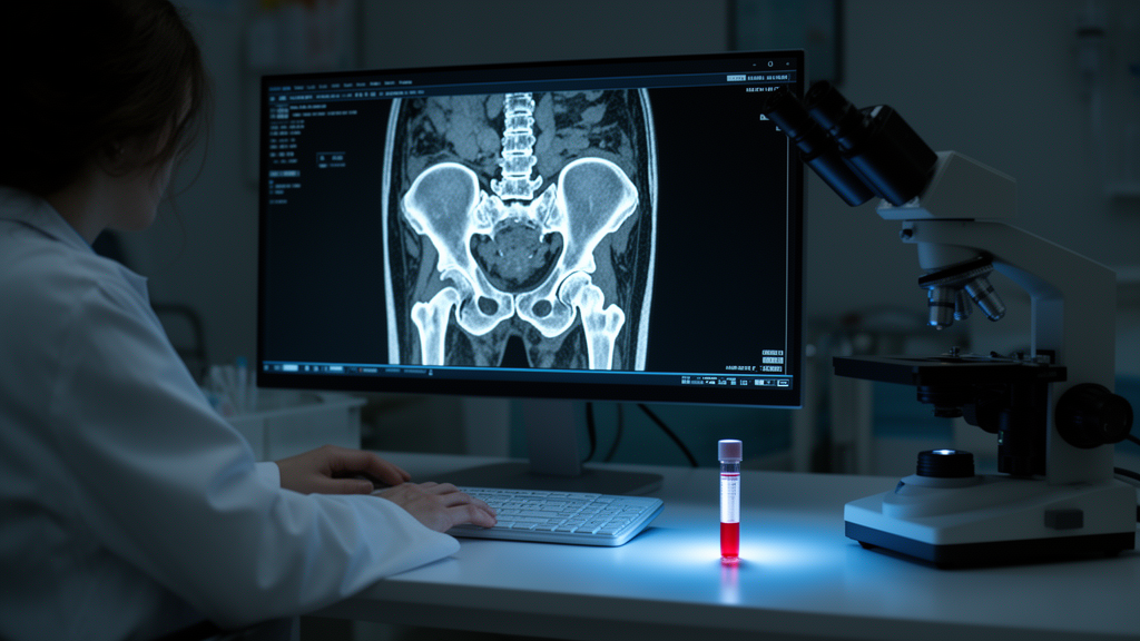
The research, led by Tao Song, Yiwen Wang, Jieni Ding, Huafeng Shou, Yunwang Chen, Hongzhi Zhang, and senior author Hong’en Xu across hospitals affiliated with Hangzhou Medical College, Zhejiang University School of Medicine, and other institutions in eastern China, set out to build a predictive tool that combines clinical information with quantitative image features extracted from computed tomography. The team focused on a specific anatomic landmark: the psoas muscle at the level of the third lumbar vertebra. This large muscle flanking the spine is a common proxy for whole-body muscle status in imaging research, and the investigators reasoned that its texture, shape, and density on CT might encode information about a patient’s physiological reserve, including how well her bone marrow and immune system would tolerate the assault of radiation and chemotherapy.
The technical foundation of the study is radiomics, a field that converts medical images into high-dimensional data. Rather than asking a radiologist to eyeball the scan, radiomics pipelines extract thousands of numerical features automatically: first-order statistics describing the distribution of pixel intensities, texture metrics capturing patterns of spatial heterogeneity, and shape descriptors quantifying geometry. In this study, the team analyzed 2,153 such features from the planning CT scans of 150 patients with surgically treated stage IB-IIA cervical cancer. All patients subsequently received postoperative radiotherapy or chemoradiotherapy, and the researchers tracked which of them developed moderate-to-severe leukopenia as an adverse event.
Extracting 2,153 features from 150 patients creates a classic statistical dilemma: far more variables than outcomes, a situation that guarantees overfitting if naive modeling is applied. The Chinese team addressed this with least absolute shrinkage and selection operator regression, universally abbreviated as LASSO, a machine learning technique that performs feature selection and regularization simultaneously. LASSO works by penalizing the absolute size of regression coefficients, effectively shrinking most of them to zero and retaining only the features that genuinely contribute predictive power. Through this process, the 2,153 candidate features were distilled down to just 10 radiomic features, which were then combined into a single composite metric called the radiomics score, or rad-score.
In parallel, the investigators examined 18 clinical variables, ranging from demographic characteristics and body mass index to treatment details and nutritional status. Multivariable analysis identified two independent clinical risk factors for moderate-to-severe leukopenia: the score on the Nutritional Risk Screening 2002 tool, a validated questionnaire used to gauge malnutrition risk in hospitalized patients, and receipt of chemotherapy as part of the adjuvant regimen. The NRS-2002 finding aligns with a growing body of evidence linking nutritional reserve to treatment tolerance, while the chemotherapy association reflects the added myelosuppressive burden that cytotoxic drugs impose on top of radiation.
The performance of the resulting models tells a nuanced story. In the training cohort of 106 patients, the rad-score alone achieved an area under the receiver operating characteristic curve, or AUC, of 0.743, while the clinical model reached 0.753. Neither was sufficient on its own. But when the radiomic signature and the clinical variables were merged into a nomogram, a graphical scoring tool that translates multiple inputs into a single probability estimate, the combined model achieved an AUC of 0.830 in the training cohort. In the internal validation cohort of 44 patients, the nomogram maintained an AUC of 0.800 with a 95 percent confidence interval of 0.641 to 0.908, while the clinical model alone dropped to 0.586 and the rad-score to 0.664. The pattern is consistent: image-derived features and clinical factors capture complementary information, and the fusion of the two outperforms either component.
An AUC of 0.8 in validation means the nomogram distinguishes patients who will develop moderate-to-severe leukopenia from those who will not with reasonable, though not perfect, discrimination. For clinicians, such a tool could change the calculus of adjuvant therapy planning. Patients flagged as high risk might receive closer white blood cell monitoring, prophylactic interventions, or modified chemotherapy scheduling, while low-risk patients could proceed with standard regimens and fewer unnecessary blood draws. The nomogram format is particularly appealing for bedside use because it requires no software: a clinician plots each variable on a scale, sums the points, and reads off the predicted probability.
The study’s authors are careful to frame their findings as hypothesis-generating rather than practice-changing. The research was single-center and retrospective, meaning the data came from existing records rather than a prospectively designed trial. The cohort of 150 patients was randomly divided into a training set of 106 and an internal validation set of 44 using a single random split, a design that tests the model on unseen data from the same institution but says nothing about whether the model generalizes to patients at other hospitals with different scanners, protocols, and populations. Radiomic features are notoriously sensitive to variations in CT acquisition parameters, reconstruction kernels, and slice thickness, which is why independent multicenter validation is considered the essential next step before any radiomics nomogram enters routine clinical use.
Nevertheless, the study adds to a rapidly expanding literature on predictive medicine, in which machine learning applied to routine imaging data generates individualized forecasts of treatment outcomes and adverse events. The idea that a scan acquired for radiation planning, before a single beam is delivered, could forecast a hematologic toxicity weeks later is a striking example of how much diagnostic information routine images contain beyond their original purpose. If multicenter validation confirms the Hangzhou team’s results, CT-based radiomics could become a standard component of pretreatment risk stratification in cervical cancer, helping oncologists anticipate leukopenia rather than react to it. For now, the nomogram remains a promising research tool, one that hints at a future where the pixels of a planning scan serve as a crystal ball for treatment tolerance.
Subject of Research: Radiomics and machine learning for predicting radiotherapy toxicity in cervical cancer
Article Title: CT-based radiomics nomogram for predicting leukopenia in surgically treated stage IB-IIA cervical cancer following (chemo) radiotherapy
Article References: Song, T., Wang, Y., Ding, J., Shou, H., Chen, Y., Zhang, H., & Xu, H. (2026). CT-based radiomics nomogram for predicting leukopenia in surgically treated stage IB-IIA cervical cancer following (chemo) radiotherapy. BMC Cancer. https://doi.org/10.1186/s12885-026-17086-9
Image Credits: AI Generated
DOI: 10.1186/s12885-026-17086-9
Keywords: radiomics, cervical cancer, leukopenia, nomogram, radiotherapy, machine learning, predictive medicine, CT-based, predicting, surgically, treated, stage
News Source: Nathaniel Bowman. (October 19, 2026). CT Scans May Predict Who Develops Leukopenia After Cervical Cancer Radiotherapy. Scienmag.