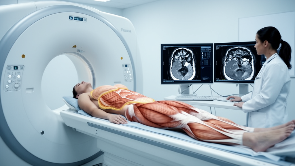
People who later develop pancreatic ductal adenocarcinoma (PDAC) show steeper declines in visceral and subcutaneous fat than matched patients without pancreatic cancer during the three years before diagnosis, according to a peer-reviewed Mayo Clinic study published in Pancreatology. The analysis also found that differences in skeletal muscle were most apparent during the final 12 months before diagnosis. Together, the findings indicate that routinely acquired CT scans may capture measurable body-composition changes associated with pancreatic cancer before it is clinically detected.
The researchers analyzed abdominal CT scans from 1,825 patients with PDAC and 3,650 matched controls from Mayo Clinic sites in Arizona, Florida and Minnesota. They used an artificial intelligence-based body-composition tool to measure changes in skeletal muscle, subcutaneous fat and visceral fat over time. By comparing longitudinal measurements between patients who developed pancreatic cancer and similar individuals without cancer, the investigators assessed whether the timing and rate of body-composition changes differed before diagnosis. The study is observational, so it shows an association between prediagnostic changes and subsequent PDAC rather than proving that those changes cause the disease or that CT-based measurements can screen for it.
The strongest observed difference was a significantly steeper decline in both subcutaneous and visceral fat during the three years before PDAC diagnosis among patients who developed the disease. Muscle differences were less apparent over the full period but became most pronounced in the year immediately preceding diagnosis. When the researchers evaluated whether the muscle and fat measurements could distinguish patients who later developed pancreatic cancer from matched controls, the measures demonstrated moderate discriminatory ability. This supports their potential to provide additional information in future risk-assessment strategies, but the study did not establish a complete clinical prediction model, evaluate a prospective screening program or demonstrate improved patient outcomes.
The results may be relevant to efforts to identify pancreatic cancer earlier because PDAC can develop silently and is often diagnosed after it has spread. Current surveillance programs focus primarily on people with inherited genetic risk or strong family histories, while most pancreatic cancers occur outside those high-risk groups. The Mayo Clinic analysis is, to the researchers’ knowledge, the largest controlled study to examine prediagnostic body-composition trajectories in patients who later developed pancreatic cancer and one of the first to assess whether those trajectories can help distinguish future cases from controls. Mayo researchers are now developing algorithms that combine body-composition measurements with other routinely collected clinical factors and imaging biomarkers to identify patients at increased risk.
Subject of Research: Prediagnostic CT body-composition trajectories associated with pancreatic ductal adenocarcinoma
Article Title: Body composition changes on CT scans may help identify pancreatic cancer risk
Article References: Body composition changes on CT scans may help identify pancreatic cancer risk. Original publication (Bibliographic details unavailable.)
Image Credits: AI Generated
DOI: Not provided
Keywords: pancreatic cancer, pancreatic ductal adenocarcinoma, body composition, CT scans, visceral fat, subcutaneous fat, skeletal muscle, artificial intelligence, early detection, Mayo Clinic, Body, composition
News Source: Nathaniel Bowman. (October 20, 2026). CT body-composition changes precede pancreatic cancer diagnosis. Scienmag.