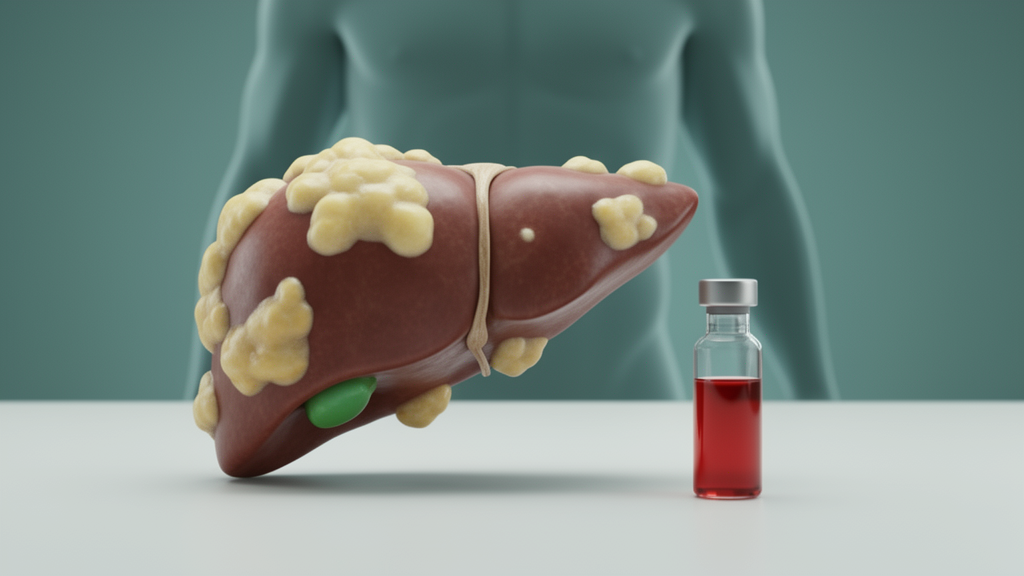
A simple blood-and-scale calculation that researchers hoped could flag fatty liver disease has stumbled in its first careful test in Egypt. In a cross-sectional study of 190 non-elderly Egyptian patients, a low creatinine-to-body weight ratio was linked to metabolic dysfunction and to steatosis defined by the Hepatic Steatosis Index, but that link dissolved once body mass index and other metabolic factors were taken into account. Only BMI remained independently associated with HSI-defined steatosis after adjustment. The findings, published in the Egyptian Liver Journal, suggest the ratio may primarily reflect body composition and obesity-related metabolic abnormalities rather than serving as an independent marker of fat in the liver.
The creatinine-to-body weight (Cre/BW) ratio is calculated by dividing serum creatinine in milligrams per deciliter by body weight in kilograms. Serum creatinine is produced largely by skeletal muscle, so in people with normal kidney function the ratio has been proposed as a cheap, easily obtainable surrogate for relative muscle mass. Because low muscle mass relative to body weight has been tied to insulin resistance, metabolic syndrome and hepatic steatosis, researchers have wondered whether the ratio could serve as a quick screening signal for metabolic dysfunction-associated steatotic liver disease, or MASLD, the condition formerly known as non-alcoholic fatty liver disease.
The rationale rests on what researchers call the muscle-liver axis. Reduced muscle mass or impaired muscle quality may contribute to insulin resistance, chronic inflammation and altered lipid metabolism, mechanisms considered key to the progression of hepatic steatosis. Serum creatinine alone, however, is influenced by absolute body size and absolute muscle mass, which is why dividing it by body weight was thought to better capture obesity-related changes in body composition. Previous studies, including a longitudinal population-based study by Okamura and colleagues and work by Lin and colleagues in non-elderly individuals, reported associations between lower Cre/BW ratios and NAFLD or metabolic abnormalities, and more recently Sun and colleagues reported an independent association between a lower ratio and CAP-defined steatosis severity in patients with MASLD.
Against that backdrop, the new study set out to evaluate the association between the Cre/BW ratio and metabolic parameters, HSI-defined steatosis and hepatic indices in non-elderly Egyptian patients, and to assess its diagnostic performance. The work was conducted at the Tropical Medicine Outpatient Clinic of the Faculty of Medicine, Tanta University, between April and August 2024, with ethical approval and written informed consent from all participants. Eligible participants were adults aged 18 to 60 years with imaging-confirmed hepatic steatosis on ultrasound or fibroscan plus at least one cardiometabolic risk factor such as obesity, diabetes, dyslipidemia, hypertension or insulin resistance. Patients with chronic kidney disease, conditions affecting muscle mass such as malignancy or rheumatologic disease, pregnancy, alcohol consumption, or viral or autoimmune hepatitis were excluded.
MASLD was defined non-invasively, combining imaging evidence of hepatic steatosis with cardiometabolic risk factors; liver biopsy was not performed because of its invasive nature, so the definition reflects a pragmatic, real-world clinical approach rather than histological confirmation. Participants were recruited consecutively from clinic attendees until the required sample size was reached, without selective sampling. Each patient underwent a detailed clinical evaluation with anthropometric measurements including BMI and waist circumference, and fasting blood tests covering complete blood count, serum creatinine, liver enzymes, lipid profile and viral hepatitis markers. Transient elastography was performed with a FibroScan 502 and three portable Echosens Mini Systems, yielding liver stiffness measurements in kilopascals and controlled attenuation parameter (CAP) values in decibels per meter, with steatosis graded S0 through S3 and fibrosis staged F0 through F4 using established cutoffs.
The resulting cohort had a mean age of 46.2 years, was 54.2 percent female, and carried a heavy metabolic burden: 43.2 percent had type 2 diabetes, 31.1 percent hypertension and 29 percent were current smokers. Most patients were overweight or obese, with a mean BMI of 34.3 kg/m2 and a mean waist circumference of 103.1 cm. Hepatic steatosis was predominantly moderate to severe, with a mean CAP of 294.6 dB/m and 52.1 percent of patients in the S3 grade, while liver stiffness values of 6.1 kPa and a FIB-4 index of 1.22 indicated generally mild fibrosis. Liver enzymes were only mildly elevated or near normal. The mean Cre/BW ratio was 0.010, and 81 percent of participants had an HSI of 36 or above, meeting the predefined criterion for HSI-defined steatosis.
The unadjusted results initially looked promising for the ratio. Patients with HSI of 36 or higher had a significantly lower Cre/BW ratio than those below the threshold, 0.009 versus 0.013. The ratio was inversely correlated with HSI, body weight, BMI and waist circumference, and positively correlated with serum creatinine, consistent with its biological basis in skeletal muscle. Notably, however, it showed no significant differences across CAP-defined steatosis grades or fibroscan-based fibrosis stages, and no significant correlations with CAP, fibroscan measurements, the FIB-4 index or liver enzyme levels. In other words, the ratio tracked anthropometric and metabolic characteristics far more closely than it tracked imaging-based measures of hepatic fat or fibrosis severity.
The diagnostic numbers told a similar story of limited usefulness. Receiver operating characteristic analysis using a Cre/BW cutoff of 0.009 or less to discriminate HSI-defined steatosis yielded a sensitivity of only 42.86 percent, meaning most true cases would be missed, although specificity was relatively high at 86.11 percent. The positive predictive value was 93.0 percent, the negative predictive value a weak 26.1 percent, and overall diagnostic accuracy 71.2 percent. The authors conclude that the limited sensitivity means the ratio would not be suitable as a standalone screening marker for HSI-defined steatosis, even though a low value was associated with the outcome in unadjusted analysis.
The decisive test came from multivariable logistic regression adjusted for age, sex, BMI, diabetes mellitus and waist circumference. The authors also flag a methodological caveat: because HSI incorporates BMI while the Cre/BW ratio contains body weight in its denominator, mathematical coupling between predictor and outcome was considered when interpreting the findings, and the observed association may partly reflect shared anthropometric and metabolic components rather than a direct biological relationship with hepatic fat accumulation.
Smokers in the cohort had slightly lower Cre/BW ratios than non-smokers, but the authors urge caution, noting that smoking status was not independently associated with the outcome in the multivariable analysis and the finding may represent chance or residual confounding. The study’s limitations are substantial: a cross-sectional, single-center design that limits causal inference and generalizability, no direct assessment of muscle mass to validate the ratio as a muscle-related marker, and reliance on HSI as the primary analytical outcome with its potential coupling problem. The authors suggest the ratio’s simplicity and low cost may still make it useful as an adjunctive marker in metabolic risk assessment, but its clinical utility for identifying steatosis requires validation in larger prospective studies using independent measures of both hepatic steatosis and body composition. For now, the message is that a number easily derived from routine labs and a weighing scale mirrors obesity and metabolic risk, but it does not stand on its own as a window into the liver.
Subject of Research: The diagnostic value of the creatinine-to-body weight ratio for metabolic dysfunction and hepatic steatosis in Egyptian patients
Article Title: Creatinine-to-body weight ratio as a marker of metabolic dysfunction and obesity-related steatosis in Egyptian patients
Article References: Ebrahim, S., Shady, E., & Mansour, S. (2026). Creatinine-to-body weight ratio as a marker of metabolic dysfunction and obesity-related steatosis in Egyptian patients. Egyptian Liver Journal, 16(1), Article 71. https://doi.org/10.1186/s43066-026-00542-3
Image Credits: AI Generated
DOI: 10.1186/s43066-026-00542-3
Keywords: MASLD, hepatic steatosis, creatinine-to-body weight ratio, Hepatic Steatosis Index, body composition, obesity, Egypt, cross-sectional study, FibroScan, BMI, muscle mass, non-invasive markers
News Source: Ophelia Keating. (October 29, 2026). Creatinine-to-body weight ratio falls short as fatty liver marker. Scienmag.