Interventional radiologists sometimes need to shut down a sizable artery with a device that stays exactly where it is placed. A study now published in CVIR Endovascular reports that a modified embolization approach, the preloading coil-in-plug technique, achieved technical success in every one of 40 treated arteries and produced no detectable recanalization, device migration, or reintervention during a mean imaging follow-up of 619 days. The retrospective single-center series, led by Takahiro Nakai and colleagues, covered celiac, internal iliac, gluteal, and external iliac arteries, making it one of the larger clinical collections of this technique and one of the few to span multiple vascular territories.
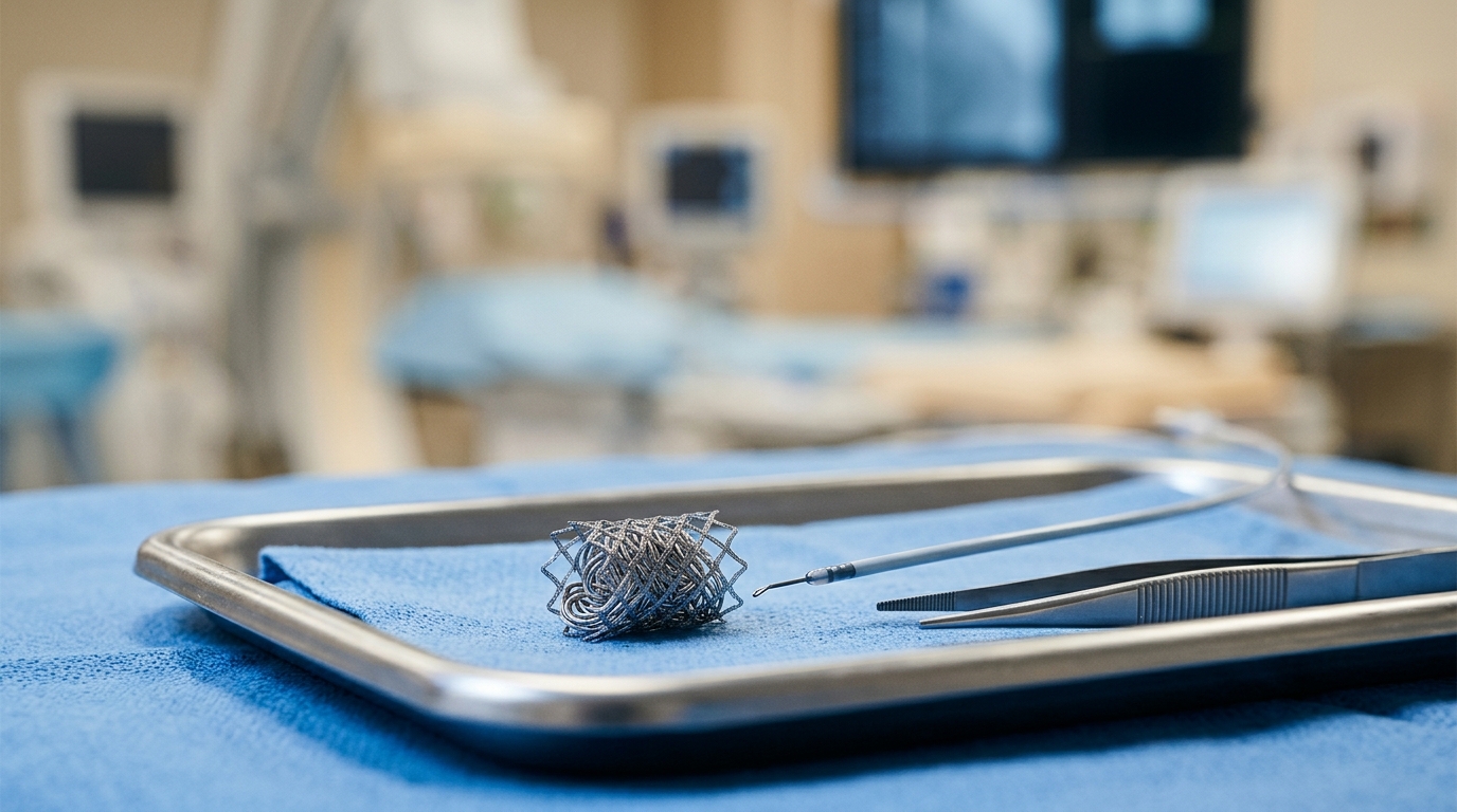
The technique builds on the Amplatzer Vascular Plug, a nitinol mesh occluder that interventionalists can deploy and reposition before releasing it. Conventional coil embolization of large or high-flow arteries may demand multiple coils and a long embolization segment, and loose coils can migrate or leave channels for persistent flow. The plug alone solves the stability problem, but complete occlusion depends partly on thrombus forming inside and around the device, so residual flow or later recanalization can occur, particularly in high-flow vessels or vessels with irregular luminal morphology. Recanalization after embolization with the single-segment plug has also been reported, although robust comparative evidence among plug models remains limited.
The coil-in-plug idea combines the two tools. In the original version, a microcatheter is advanced through the nitinol mesh into a plug already deployed inside the target vessel, and coils are then packed into the expanded device before it is detached. That cannulation step can be technically challenging. The preloading variant simplifies it: before insertion, the operator passes a microcatheter through the nitinol mesh into the internal space of the plug outside the patient, loads plug and microcatheter together into a guiding sheath or catheter, advances them to the target, and deploys the plug at the intended site without immediate detachment. Detachable coils are then delivered through the preloaded microcatheter and packed within the expanded plug. The plug acts as a scaffold that limits coil migration, while the internal packing increases the amount of embolic material and reduces residual flow channels through the mesh.
The study team reviewed their institutional interventional radiology database for consecutive patients treated with this technique between April 2018 and April 2026. Patients qualified when a first-generation Amplatzer plug was placed in a target artery and one or more metallic coils were intentionally deployed within it before detachment. Thirty-four patients, 21 of them men with a mean age of 77.3 years, contributed 40 embolized vessels. Twenty-nine patients had one vessel treated, four had two, and one had three. The targets were the celiac artery in six cases, the proximal internal iliac artery in 22, the distal internal iliac artery in three, the superior or inferior gluteal artery in eight, and the external iliac artery in one. Every treated vessel was part of a combined strategy with proximal stentgraft implantation.
Procedurally, the plug was generally sized about 30 to 50 percent larger than the measured target-vessel diameter, and coils were chosen with a nominal diameter roughly equal to the vessel. The plug was detached only after the team confirmed satisfactory device position, absence of clinically significant coil protrusion, and adequate reduction or cessation of flow. The mean plug-to-vessel diameter ratio came out at 1.44, corresponding to 44 percent oversizing, which the authors note sits within the generally recommended range for plug embolization. On average, 3.13 coils were packed within each plug, and the mean coil-packing time, measured from the angiographic record immediately before or after plug deployment to the acquisition taken just before detachment, was 10.7 minutes. The authors suggest this shows internal packing can generally be completed within a clinically acceptable period.
Technical success, defined as successful plug placement, successful coil deployment within it, and satisfactory angiographic occlusion of the target vessel at the end of the procedure, was achieved in all 40 vessels, and satisfactory final angiographic occlusion was achieved in all of them. In 10 vessels, adjacent branches outside the plug-covered segment were also embolized. No procedure-related complication occurred during the procedure or follow-up: no non-target embolization, clinically significant ischemia, device migration, vessel injury, or embolization-related reintervention. Ten patients died during the observation period, all of whom had undergone single-vessel embolization, but none of the deaths was related to the embolization procedure or to ischemia of the treated territory; the causes included cancer, pneumonia, cardiac death, chronic renal failure, retroperitoneal bleeding, and sepsis.
Follow-up relied on contrast-enhanced computed tomography, generally performed at about one week, six months, and one year after treatment and every six to twelve months thereafter, with timing adjusted to each patient’s condition. The mean imaging follow-up was 619 days, with a median of 367 days and a range of 3 to 1826 days. No target-vessel recanalization, persistent flow requiring treatment, or device migration appeared in either of two hemodynamically distinct configurations: 29 vessels in which the plug sat adjacent to the stentgraft, and 11 in which an internal iliac artery aneurysm sac was interposed between the stentgraft and the treated branch. For the 10 patients who died, the final scan before death still showed maintained occlusion of the treated vessel.
The authors are careful about what these numbers can and cannot show. Because all target vessels were treated alongside proximal stentgraft implantation, and because the two configurations differ hemodynamically, maintenance of distal branch occlusion cannot be attributed to the coil-in-plug technique alone; the stratified findings should be read as imaging outcomes of the combined treatment rather than a direct comparison of the technique’s intrinsic performance across territories. With zero recanalization events among 40 vessels, the rule of three gives an approximate upper 95 percent confidence bound of 7.5 percent for the underlying rate, so the true event rate cannot be assumed to be zero. Metallic coils can also produce CT artifacts that may obscure subtle residual or recurrent contrast opacification, reducing the sensitivity of CT-based assessment compared with plug-alone embolization.
Other limitations shape the interpretation. The study was retrospective and single-center with no control group, so the relative advantages over plug placement alone or conventional coil embolization could not be determined. Vessels and indications were heterogeneous, five patients contributed more than one vessel so the observations were not fully independent, follow-up duration varied, and death shortened surveillance in 10 patients. Rare late mechanical complications cannot be excluded; delayed problems such as plug erosion into adjacent structures have been reported elsewhere, and the authors call for longer-term surveillance. Immediate angiographic occlusion was also not graded with a prospectively defined system, and future studies should use standardized criteria to test whether residual flow right after plug detachment predicts later recanalization.
Even with those caveats, the series offers a useful observational signal. The mean coil count of 3.1 per plug was comparable to values of 3.8 and 3.36 reported in earlier studies of conventional and preloading coil-in-plug techniques, and earlier comparative work found those approaches used fewer coils and shorter embolization lengths than conventional coil embolization. Newer devices, including shape-memory polymer plugs and low-profile membrane-covered occluders, are expanding the options for short-segment embolization, but direct comparative data with the preloading coil-in-plug technique remain limited. The authors conclude that the technique is a feasible option for controlled short-segment arterial embolization and that further comparative studies should clarify optimal plug oversizing and coil packing, and determine whether it truly outperforms conventional coils or a plug alone.
Subject of Research: Mid-term outcomes of the preloading coil-in-plug embolization technique across multiple arterial territories
Article Title: Clinical outcomes and midterm durability of the preloading coil-in-plug technique in various arterial territories
Article References: Nakai, T., Iwakoshi, S., Iwata, M., Kishida, H., Oshima, K., Ichihashi, S., & Tanaka, T. (2026). Clinical outcomes and midterm durability of the preloading coil-in-plug technique in various arterial territories. CVIR Endovascular, 9(1), Article 139. https://doi.org/10.1186/s42155-026-00790-9
Image Credits: AI Generated
DOI: 10.1186/s42155-026-00790-9
Keywords: embolization, Amplatzer Vascular Plug, coil-in-plug technique, interventional radiology, recanalization, stentgraft, internal iliac artery, celiac artery, vascular occlusion, contrast-enhanced CT, mid-term durability, Clinical
News Source: Ophelia Keating. (November 5, 2026). Coil-packed plug technique shows durable artery occlusion across territories. Scienmag.