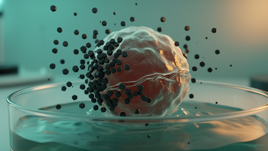
Strontium hexaferrite (SrFe12O19) nanoparticles, a chemically robust hard-magnetic material with high magnetic anisotropy, have been engineered as a cobalt-free platform for non-thermal magneto-mechanical cancer therapy, according to a study published in the Journal of Nanoparticle Research. In laboratory tests on 4T1 murine adenocarcinoma cells, a pronounced magneto-mechanical effect was observed when the nanoparticles were exposed to a low-frequency alternating magnetic field of 100 mT at 22 Hz: cell viability fell from approximately 55% to approximately 25% at a concentration of 200 µg/mL, with an IC50 of roughly 150 µg/mL. The nanoparticles themselves showed low intrinsic cytotoxicity at concentrations up to 50 µg/mL.
The research team, led by T. R. Nizamov and colleagues, synthesized the particles via a citrate precursor route followed by rapid annealing at 950 °C for either 5 or 30 minutes. The particles were then subjected to high-energy planetary ball milling in an oleic-acid/octadecene medium for up to 48 hours, and finally functionalized with PEG to enable dispersion in water. The study systematically correlated processing-dependent changes in phase composition, crystallite size, lattice parameters, magnetic properties, and colloidal behavior with biological performance.
X-ray diffraction confirmed predominantly single-phase SrFe12O19 after annealing, although prolonged milling proved to be a decisive processing step. Extended milling eliminated residual hematite, reduced the crystallite size from roughly 23–25 nm to approximately 8–12 nm, and induced lattice expansion and partial amorphization. These structural changes came at a magnetic cost: coercivity, remanence, and saturation magnetization all decreased as milling progressed.
PEGylation produced stable aqueous dispersions with hydrodynamic diameters of approximately 165–175 nm and low polydispersity, an important characteristic for any material intended for biomedical use. However, the magnetic response was further reduced after functionalization, an effect the authors attribute to the non-magnetic polymer shell and to size-selective fractionation during processing.
The strongest biological effect was observed for the nanoparticles that had been annealed for 30 minutes and then milled for 48 hours, identifying that processing combination as the most effective configuration tested.
The findings are limited to in vitro experiments on a murine adenocarcinoma cell line, and the reported viability reductions reflect cell culture measurements rather than clinical outcomes. Nevertheless, the study links each stage of materials processing—from annealing time and milling duration to polymer coating—to measurable changes in both magnetic behavior and biological effect, offering a detailed processing-performance map for this class of hard-magnetic nanoparticles.
Subject of Research: Synthesis and magneto-mechanical cancer therapy potential of strontium hexaferrite nanoparticles
Article Title: Synthesis, structural evolution, and magneto-mechanical effect of SrFe12O19 nanoparticles for biomedical applications
Article References: Nizamov, T. R., Bordyuzhin, I. G., Ivanova, A. V., Abakumov, M. A., Shchetinin, I. V., & Savchenko, A. G. (2026). Synthesis, structural evolution, and magneto-mechanical effect of SrFe12O19 nanoparticles for biomedical applications. Journal of Nanoparticle Research, 28(11), Article 277. https://doi.org/10.1007/s11051-026-06799-4
Image Credits: AI Generated
DOI: 10.1007/s11051-026-06799-4
Keywords: strontium hexaferrite, SrFe12O19 nanoparticles, magneto-mechanical therapy, alternating magnetic field, 4T1 cancer cells, PEGylation, ball milling, coercivity, crystallite size, biomedical applications, cobalt-free, Journal of Nanoparticle Research
News Source: Nathaniel Bowman. (October 28, 2026). Cobalt-Free Strontium Hexaferrite Nanoparticles Show Magneto-Mechanical Cancer Cell Killing in Vitro. Scienmag.