A cell-based study has identified a molecular link between the copper-carrying protein ceruloplasmin, abbreviated CP, and cuproptosis-associated changes in castration-resistant prostate cancer cells. Researchers combined analysis of 550 prostate cancer tumors and 52 normal tissues from the TCGA-PRAD dataset with experiments in two androgen-independent cell lines, PC-3 and 22Rv1. Reducing CP in 22Rv1 cells increased the expression of FDX1, DLAT and lipoylated proteins, raised mitochondrial reactive oxygen species and inhibited proliferation. Restoring the long non-coding RNA STPG3-AS1 reversed those changes, supporting a CP-STPG3-AS1 regulatory axis. The results indicate a cuproptosis-sensitized state rather than proving that cuproptosis itself had been completed, and the work remains preclinical.
The study focused on an unresolved question in prostate cancer biology. CP is a copper-binding glycoprotein involved in copper transport and homeostasis, while cuproptosis is a copper-dependent form of regulated cell death associated with lipoylated tricarboxylic acid cycle proteins. Earlier studies had linked copper metabolism with prostate cancer and had reported that several established cuproptosis regulators, including FDX1, are elevated in tumors. Most proposed connections between CP and cuproptosis in prostate cancer, however, relied on computational correlations or observational gene-expression patterns rather than functional experiments with knockdown and rescue.
To explore the relationship, the researchers assembled a list of 1,046 genes annotated as cuproptosis-related across several databases and published collections. They compared this list with CP-associated genes, identifying an intersection of 80 genes. Among the intersection set, 58 were downregulated and 22 were upregulated in prostate cancer relative to normal tissue. Functional annotations linked the set to copper-ion homeostasis, copper binding, cell-death regulation and oxidative stress. A protein-interaction network identified ATP7A, ATP7B, SOD1, SOD2, HMOX1 and other copper-handling or stress-response proteins as highly connected nodes, although the network alone could not establish causal relationships.
Expression analysis produced a distinction between CP and the core cuproptosis machinery. The transcript levels of FDX1, DLST, LIAS and DLD were each significantly higher in prostate cancer tissues than in normal samples, with all reported P values below 0.001. CP itself did not differ significantly between the groups, with P equal to 0.46. CP was nevertheless weakly correlated with the four core genes in tumor tissue: the correlation coefficients ranged from 0.205 for FDX1 to 0.300 for DLST. These modest associations suggest co-expression, not direct regulation, and survival analyses found no independent prognostic value for CP, FDX1, DLST, LIAS or DLD. No significant overall-survival difference was detected between groups divided at the median expression of any of these genes.
In PC-3 cells, which represent an androgen-receptor-null form of castration resistance, the team added purified human serum CP at concentrations of 50 or 100 micrograms per milliliter for 24 or 48 hours. The CCK-8 assay, which measures metabolic activity associated with cellular dehydrogenase activity rather than cell number directly, showed a concentration- and time-dependent reduction. Relative metabolic activity at 100 micrograms per milliliter was 88.08% of control after 24 hours and 91.26% after 48 hours. The effect was mild. Hoechst staining also found more cells with nuclear condensation or fragmentation, rising from 9% to 14% in controls to 22% to 31% at the highest concentration. These observations show that CP exposure was associated with reduced metabolic activity and apoptosis-related morphology, but they do not by themselves identify the mechanism or establish a clinically useful effect.
The more detailed perturbation experiments were performed in 22Rv1 cells, an androgen-independent line that expresses full-length androgen receptor and the splice variant AR-V7. CP knockdown reduced STPG3-AS1 expression by at least 30%, while the protein measurements showed increases of at least 50% in FDX1, DLAT and lipoylated proteins. The mitochondrial reactive oxygen species signal measured by MitoSOX flow cytometry rose by at least 40% in the CP-silenced group, and CCK-8 analysis showed proliferation inhibition of at least 30% at 72 hours. These are cuproptosis-associated molecular changes, defined in the study by increased lipoylation machinery, lipoylated proteins and mitochondrial oxidative stress; they are not direct evidence of completed cuproptotic death.
The rescue experiment strengthened the proposed mediation. Co-expression of STPG3-AS1 after CP knockdown brought the elevated FDX1, DLAT and lipoylated protein measurements back to within 80% of control levels, reduced the mitochondrial reactive oxygen species signal to within 75% of control and reduced proliferation inhibition to no more than 15%. In that sense, STPG3-AS1 was necessary for the measured consequences of CP depletion and sufficient, at the tested expression level, to reverse them. The authors also perturbed the copper importer SLC31A1, which encodes CTR1. This manipulation reduced LIPE-AS1 and AC005790.1 expression and produced parallel changes in the same experimental readouts, with LIPE-AS1 overexpression reversing those effects. That comparison was included as a specificity control for copper-homeostasis pathways, not as evidence that the two long non-coding RNAs are equivalent.
Several technical constraints prevent the findings from being interpreted as a demonstration of cuproptosis in patients. The experiments did not measure intracellular labile copper, perform copper-chelator rescue, establish FDX1 dependence, demonstrate DLAT oligomerization or show loss of iron-sulfur cluster proteins, all of which the authors identify as requirements for more definitive cuproptosis classification. The study also used only two established CRPC cell lines. It included no primary prostate cancer cells, androgen-dependent cell lines, animals or clinical intervention, and its survival analysis relied on one public dataset without external validation or adjustment for clinical covariates. STPG3-AS1’s direct molecular targets also remain unknown; the proposed mechanisms include microRNA competition, transcriptional regulation and other RNA-mediated effects, but none was tested in the reported experiments.
Overall, the work provides a testable model in which CP restrains lipoylation-related and cuproptosis-associated changes in 22Rv1 cells, partly through STPG3-AS1. The contrast between the two models is also informative: adding CP to PC-3 cells produced apoptosis-like nuclear changes, whereas reducing CP in 22Rv1 cells increased markers associated with cuproptosis sensitization. The results do not establish that CP is a therapeutic target, that STPG3-AS1 can safely be manipulated in people or that targeting the pathway would improve survival. Instead, they identify a mechanism requiring direct copper measurement, stricter death-pathway validation, broader cell models, animal studies and appropriately controlled clinical cohorts before any clinical inference can be made.
Subject of Research: Ceruloplasmin and STPG3-AS1 regulation of lipoylation-associated changes in castration-resistant prostate cancer cells
Article Title: The CP-STPG3-AS1 axis regulates lipoylation and cuproptosis in castration-resistant prostate cancer
Article References: He, J. W., Li, P. Z., & Zhang, Z. Z. (2026). The CP-STPG3-AS1 axis regulates lipoylation and cuproptosis in castration-resistant prostate cancer. Frontiers in Oncology, 16, Article 1989277. https://doi.org/10.3389/fonc.2026.1989277
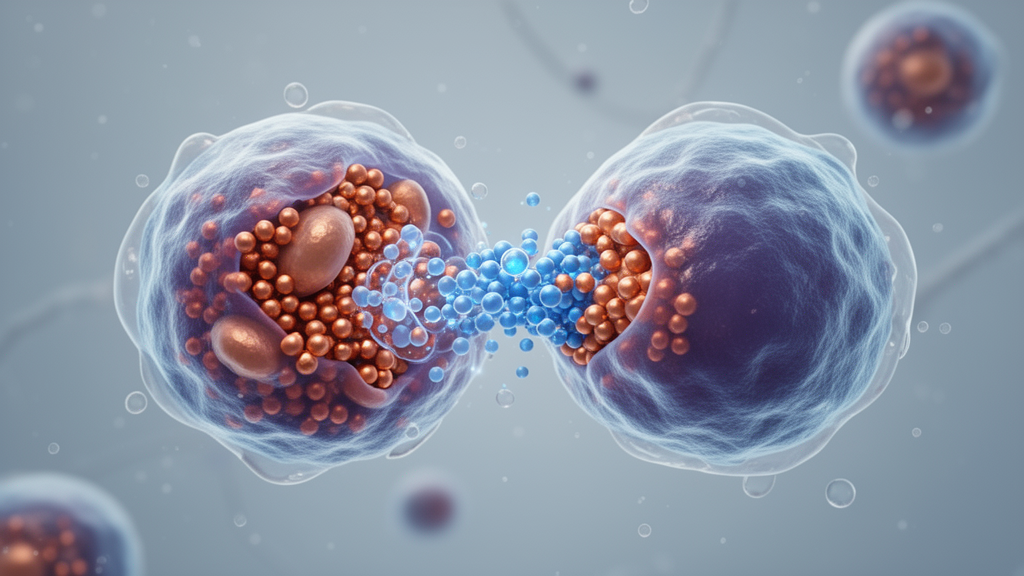
Image Credits: AI Generated
DOI: 10.3389/fonc.2026.1989277
Keywords: castration-resistant prostate cancer, ceruloplasmin, cuproptosis, STPG3-AS1, lipoylation, FDX1, mitochondrial ROS, long non-coding RNA, prostate cancer, cell culture, CP-STPG3-AS1, axis
News Source: Nathaniel Bowman. (October 22, 2026). Ceruloplasmin regulates cuproptosis-related changes in resistant prostate cancer cells. Scienmag.