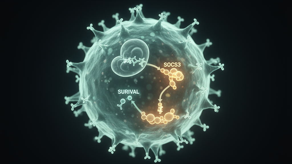
Glioblastoma remains one of the most difficult cancers to treat, and a new laboratory study now points to a molecular switch that can simultaneously slow tumor growth and push tumor cells toward self-destruction. Researchers report that suppressor of cytokine signaling 3, or SOCS3, a protein long known as a brake on the JAK-STAT signaling pathway, suppresses glioblastoma cell proliferation and drives apoptosis by activating the mitochondrial unfolded protein response, a stress program centered on the cell’s energy factories. The work, published in the journal Cell Division, combines experiments in glioblastoma cell lines with xenograft models in nude mice and identifies a mechanistic link between JAK-STAT inhibition and mitochondrial stress that could inform future therapeutic strategies.
Using reverse transcription quantitative polymerase chain reaction and Western blotting, they found that both the mRNA and protein levels of SOCS3 were significantly lower in the tumor cells than in the normal astrocytes, with A172 cells showing the more pronounced decrease. That observation fits a broader pattern: in many cancers, SOCS3 expression is frequently suppressed or functionally silenced, and in glioblastoma its loss has been linked to high malignancy, chemoresistance, and recurrence. Because A172 cells showed the strongest deficit, the researchers selected that line for most of the subsequent functional experiments.
To test what restoring the protein would do, the group engineered A172 cells to overexpress SOCS3 and, in parallel, generated knockdown lines in which the gene was silenced. The results were striking and bidirectional. Overexpression of SOCS3 significantly increased the abundance of pro-apoptotic proteins including Bax, cleaved caspase-3, and cleaved caspase-9, while reducing the anti-apoptotic protein Bcl-2. Terminal deoxynucleotidyl transferase-mediated dUTP nick-end labeling, a staining technique that flags cells undergoing apoptosis, showed a significantly higher proportion of dying cells in the SOCS3-overexpressing group. At the same time, MTT assays revealed that cell proliferation was significantly suppressed when SOCS3 was elevated, and Western blots showed reduced levels of the proliferation markers Ki67 and PCNA. Knocking SOCS3 down produced the mirror image: less apoptosis, more proliferation, and higher levels of the growth markers.
With a clear functional effect established, the researchers turned to proteomics to understand how SOCS3 achieves it. Comparing A172 cells carrying SOCS3 overexpression against controls, they identified 52 differentially expressed proteins, 33 of them upregulated and 19 downregulated. Gene Ontology and KEGG pathway analysis tied these changes to apoptosis, the unfolded protein response, mitochondrial autophagy, and the JAK-STAT pathway. Gene set enrichment analysis added a directional clue: pro-apoptotic and unfolded protein response pathways were predominantly enriched in the SOCS3-overexpressing cells, while pathways associated with mitochondrial autophagy and JAK-STAT signaling were enriched in the controls. The team then mined The Cancer Genome Atlas glioblastoma dataset and found that SOCS3 expression correlated positively with the cell senescence gene CDKN1A, the pro-apoptotic gene BAX, and the mitochondrial unfolded protein response genes ATF5 and DDIT3, and negatively with cell cycle genes CCNA2 and CDK2, the anti-apoptotic gene BCL2, and the mitophagy genes PINK1 and PRKN.
Notably, the same public data analysis produced a result that complicates any simple tumor-suppressor narrative. In Kaplan-Meier survival analyses of glioblastoma patients, high SOCS3 expression was associated with significantly worse outcomes rather than better ones. Patients with high SOCS3 expression had shorter overall survival, with a hazard ratio of 4.37 and a 95 percent confidence interval of 3.31 to 5.76, and also worse disease-specific survival, with a hazard ratio of 4.51. The progression-free interval analysis pointed in the same direction, with a hazard ratio of 2.89. The experimental work, by contrast, shows that forcing SOCS3 expression in tumor cells directly suppresses their growth, so the clinical correlation and the cellular manipulation point in different directions and the study does not resolve that tension.
To pin down which apoptotic route SOCS3 uses, the researchers examined three canonical cell death pathways: the death receptor pathway, the endoplasmic reticulum stress pathway, and the mitochondrial-dependent pathway. Western blotting found no significant differences in death receptor proteins such as Fas, FasL, TNF-alpha, and cleaved caspase-8, nor in endoplasmic reticulum stress markers including p-PERK, GRP78, cleaved caspase-7, and cleaved caspase-12, across the experimental groups. The mitochondrial route told a different story. JC-1 staining, which reports mitochondrial membrane potential through a color shift, showed that SOCS3 overexpression significantly reduced membrane potential, a hallmark of mitochondrial depolarization, while knockdown increased it. Immunofluorescence revealed that cytochrome C, a protein that escapes mitochondria to trigger the apoptotic cascade, was released into the cytoplasm at significantly higher levels in SOCS3-overexpressing cells. A caspase-9 activity assay confirmed that the initiator caspase of the intrinsic pathway was significantly more active when SOCS3 was abundant.
The mechanistic core of the paper lies in connecting this mitochondrial death program to JAK-STAT signaling. When the researchers treated SOCS3-overexpressing A172 cells with interleukin-6, a cytokine that activates JAK2 and STAT3, the mitochondrial stress response was blunted: levels of the mitochondrial chaperones and proteases HSP60, ClpP, mtHSP70, and LONP1, along with the stress-responsive transcription factor CHOP, all fell, and phosphorylation of JAK2 and STAT3 rose. TUNEL staining showed that apoptosis was significantly reduced in the SOCS3-overexpressing cells treated with interleukin-6 compared with SOCS3 overexpression alone. Conversely, when SOCS3 was knocked down, JAK2 and STAT3 phosphorylation increased and the mitochondrial stress proteins decreased, and treating those knockdown cells with ruxolitinib, a JAK2/STAT3 inhibitor, restored both the stress proteins and the apoptotic rate.
To push the stress response further, the team added nicotinamide riboside, a compound that induces mitochondrial stress, to the SOCS3-overexpressing cells that had been rescued by interleukin-6. TUNEL staining showed a significant increase in apoptosis in the triple-treated group compared with the group receiving SOCS3 overexpression and interleukin-6 alone, indicating that the mitochondrial stress pathway could be re-engaged even when JAK-STAT signaling was active. The same logic was then tested in animals. The researchers implanted A172 and U251 glioblastoma cells, either with or without SOCS3 overexpression, subcutaneously into the right flank of nude mice and, in parallel, established orthotopic intracranial models. Ten days after implantation, the mice received interleukin-6, nicotinamide riboside, or both. Tumors in the SOCS3-overexpressing groups were significantly smaller in both volume and weight than controls. Interleukin-6 reversed this suppression and increased tumor burden, while nicotinamide riboside counteracted the tumor-promoting effect of interleukin-6. Western blotting of tumor tissue showed that SOCS3 overexpression upregulated the mitochondrial stress proteins HSP60 and ClpP and the apoptosis-related proteins caspase-3 and caspase-9 while downregulating Bcl-2, and that interleukin-6 diminished these changes. TUNEL staining of tumor sections confirmed a substantial increase in apoptotic cells in the SOCS3-overexpressing groups, an effect that was mitigated by interleukin-6 and restored by nicotinamide riboside. Immunohistochemistry showed that SOCS3 overexpression also significantly reduced the proliferation markers Ki67 and PCNA in tumor tissue.
The authors frame these findings as the first demonstration that SOCS3 induces apoptosis in glioblastoma cells through the mitochondrial unfolded protein response pathway by regulating intracellular environmental stress, and they argue that the protein exerts dual tumor-suppressive effects by inhibiting JAK-STAT signaling and activating mitochondrial stress pathways. They acknowledge that the study has limitations: the in vivo work relied on A172 and U251 xenografts in nude mice, no clinical samples or patient cohorts were included for validation, and the analysis focused on the JAK-STAT and mitochondrial unfolded protein response pathways while SOCS3 may influence glioblastoma biology through other signaling networks. They also note that translating these results into therapy would face practical challenges, including tissue-specific expression, the short half-life of the SOCS3 protein, potential disruption of normal immune function, and limits on delivery efficiency and specificity. Potential strategies they discuss include gene therapy approaches using viral or nanoparticle vectors to raise SOCS3 expression in tumor tissue, or screening for compounds that promote SOCS3 expression or stabilize the protein, as well as combining SOCS3 pathway modulation with existing treatments or with immunotherapies such as CAR-T cells. For now, the work stands as a cell-line and mouse-model study that maps a specific molecular circuit, showing that a single protein can act as both a brake on a growth signaling pathway and an accelerator of a mitochondrial death program in glioblastoma cells.
Subject of Research: SOCS3 regulation of JAK-STAT signaling and the mitochondrial unfolded protein response in glioblastoma cells
Article Title: SOCS3 suppresses glioblastoma growth via JAK-STAT inhibition and mitochondrial unfolded protein response activation
Article References: Hou, G., Song, Z., Wang, J., Wang, J., Wang, H., & Li, T. (2026). SOCS3 suppresses glioblastoma growth via JAK-STAT inhibition and mitochondrial unfolded protein response activation. Cell Division, 21(1), Article 9. https://doi.org/10.1186/s13008-026-00178-0
Image Credits: AI Generated
DOI: 10.1186/s13008-026-00178-0
Keywords: glioblastoma, SOCS3, JAK-STAT, mitochondrial unfolded protein response, apoptosis, xenograft, A172, U251, interleukin-6, nicotinamide riboside, ruxolitinib, Cell Division
News Source: Nathaniel Bowman. (October 30, 2026). Cell study reveals SOCS3 as a dual brake on glioblastoma growth and survival. Scienmag.