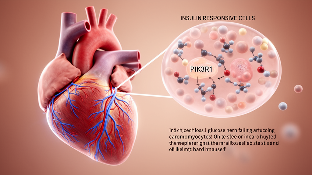
In a mouse model of diabetic cardiomyopathy, selectively deleting the PIK3R1 gene from heart muscle cells improved cardiac function, reduced iron-dependent lipid damage known as ferroptosis and preserved the balance between mitochondrial fission and fusion. The study, published in Frontiers in Cell and Developmental Biology, also found that PIK3R1 loss increased signaling through the PI3K/Akt/Nrf2 pathway. That pathway was associated with the protective effects, but the experiments did not directly establish it as the mechanism, making it a candidate mediator rather than a proven causal link.
The researchers began with public myocardial transcriptome data and found that Pik3r1 was significantly upregulated in diabetic cardiomyopathy. It was one of four genes shared among genes elevated in the disease, a curated inventory of mitochondrial genes and a set of ferroptosis-promoting genes. The same elevation appeared in three independent mouse datasets, while pathway analysis identified PI3K-Akt signaling as the most significantly enriched among upregulated pathways. These analyses supplied the rationale for testing PIK3R1 experimentally, although computational association alone could not establish a causal role.
To model diabetic cardiomyopathy, investigators fed male C57BL/6J mice a high-fat diet containing 60% fat and then administered three daily injections of low-dose streptozotocin. At week 24, model animals had elevated fasting glucose, impaired glucose tolerance, reduced left ventricular ejection fraction and fractional shortening, myocardial hypertrophy and fibrosis. Their cardiomyocytes also showed increased PIK3R1 expression. The experimental program included five cohorts of 10 mice per group, with separate tests of ferroptosis inhibition, mitochondrial fission inhibition, PI3K/Akt activation, cardiomyocyte-specific PIK3R1 knockout and a rescue involving the mitochondrial fission protein Drp1.
Two pharmacological experiments indicated that ferroptosis and excessive mitochondrial fission contribute to the phenotype in this model. Ferrostatin-1, a ferroptosis inhibitor, improved ejection fraction and fractional shortening, reduced myocardial remodeling and fibrosis, lowered serum cardiac injury markers and decreased TUNEL-positive apoptotic cells in diabetic mice. Mdivi-1, used to inhibit mitochondrial fission, produced similar improvements in cardiac performance, tissue injury and apoptosis. The study did not design an experiment to divide the relative contributions of ferroptosis, apoptosis and mitochondrial dysfunction, and it did not examine other regulated cell-death pathways.
Examination of the diabetic hearts linked altered mitochondrial dynamics with ferroptosis. Transmission electron microscopy showed atrophic mitochondria with increased membrane density, disrupted cristae and abnormal shapes. The hearts also accumulated iron, displayed elevated reactive oxygen species and had higher malondialdehyde, lower glutathione and a lower glutathione-to-oxidized-glutathione ratio. A fluorescent lipid probe shifted toward its oxidized form, while the ferroptosis-associated proteins ACSL4 and GPX4 moved in directions consistent with increased ferroptotic stress: ACSL4 increased and GPX4 decreased. After Mdivi-1 treatment, mitochondrial structure and these biochemical and molecular indicators improved together, though the reliance on one inhibitor means off-target effects cannot be excluded.
The central experiment used cardiomyocyte-specific PIK3R1 knockout mice generated with the Cre-LoxP system. Compared with wild-type diabetic mice, knockout mice had better left ventricular ejection fraction and fractional shortening, less hypertrophy and fibrosis, lower serum creatine kinase, creatine kinase-MB and lactate dehydrogenase, and fewer apoptotic cardiomyocytes. Knockout also preserved mitochondrial morphology, increased myocardial ATP content and mitochondrial DNA copy number, lowered reactive oxygen species, and shifted the balance of mitochondrial regulatory proteins toward fusion and autophagy rather than fission. Iron deposition and lipid peroxidation fell, GPX4 and SLC7A11 rose, and ACSL4 declined. These convergent measurements associate cardiomyocyte PIK3R1 deficiency with improved mitochondrial homeostasis and reduced ferroptosis in diabetic mice.
Signaling measurements followed a similar pattern. In wild-type diabetic mice, phosphorylation of PI3K and Akt decreased, as did nuclear Nrf2 and the downstream protein HO-1. The PI3K/Akt agonist 740 Y-P increased these signals and was accompanied by lower oxidative stress, higher ATP content, more active mitophagy and improved mitochondrial dynamics. PIK3R1-deficient mice showed greater pathway activation and better mitochondrial measures than corresponding wild-type groups. Because PIK3R1 encodes the p85α regulatory subunit that restrains PI3K, the increased PI3K and Akt phosphorylation after knockout was consistent with effective loss of the inhibitory subunit, although deletion efficiency was inferred rather than quantified directly in cardiac tissue.
A rescue experiment tested whether Drp1-driven mitochondrial fission could erase the cardiac protection associated with PIK3R1 loss. Researchers delivered an adeno-associated virus carrying Drp1 or an empty control vector to knockout diabetic mice. Drp1 overexpression reversed the knockout-associated improvements in ejection fraction and fractional shortening, worsened myocardial remodeling, raised serum cardiac injury markers and increased TUNEL-positive apoptosis. It also returned the expression of apoptosis-related proteins toward the diabetic wild-type pattern. These results place Drp1 downstream of PIK3R1 for cardiac function, remodeling and apoptosis. They do not show that Drp1 mediates the mitochondrial or ferroptotic effects, because those endpoints were not measured in the rescue cohort.
The findings offer a potentially useful research target, but they remain preclinical. The work included no cultured cardiomyocytes, no human myocardium and no clinical specimens, so cell autonomy and relevance to patients remain unknown. The investigators also did not disable PI3K, Akt or Nrf2 in PIK3R1-deficient animals, which would have been necessary to establish pathway dependence. Nuclear Nrf2 was evaluated by protein analysis without DNA-binding or antioxidant-response-element reporter tests, and several additional ferroptosis markers were absent. Only male mice were studied, all endpoints came from one 24-week time point, and group sizes followed comparable modeling practice rather than a prior power calculation. PIK3R1 therefore warrants further evaluation as a candidate therapeutic target, but the results do not establish a treatment for diabetic cardiomyopathy in people.
Subject of Research: Experimental mouse evidence linking cardiomyocyte PIK3R1 loss with improved mitochondrial dynamics, reduced ferroptosis and attenuated diabetic cardiomyopathy.
Article Title: Cardiomyocyte PIK3R1 deficiency attenuates mitochondrial dynamic imbalance and ferroptosis in diabetic cardiomyopathy in association with the PI3K/Akt/Nrf2 axis
Article References: Zhou, J., Xiao, Q., Zhang, F., Shen, X., Wu, S., Wang, L., & Huang, C. (2026). Cardiomyocyte PIK3R1 deficiency attenuates mitochondrial dynamic imbalance and ferroptosis in diabetic cardiomyopathy in association with the PI3K/Akt/Nrf2 axis. Frontiers in Cell and Developmental Biology, 14, Article 1953037. https://doi.org/10.3389/fcell.2026.1953037
Image Credits: AI Generated
DOI: 10.3389/fcell.2026.1953037
Keywords: PIK3R1, diabetic cardiomyopathy, ferroptosis, mitochondrial dynamics, PI3K/Akt/Nrf2, cardiomyocytes, Drp1, mice, oxidative stress, Cardiomyocyte, deficiency, attenuates
News Source: Juliet Wilcox. (October 21, 2026). Cardiomyocyte PIK3R1 Loss Protects Hearts in Diabetic Mouse Model. Scienmag.