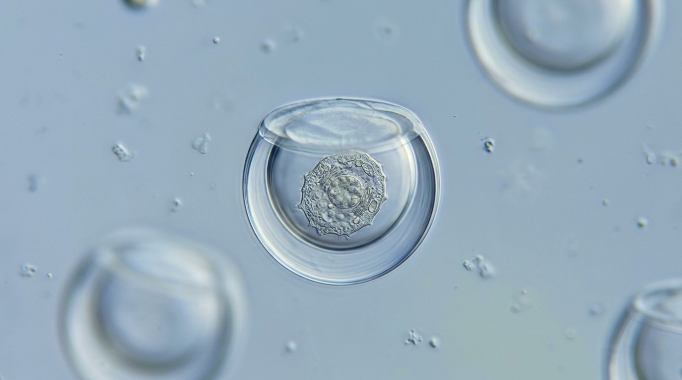
In a study published in Science Advances, bioengineers at the University of California, Los Angeles, introduced a self-assembling capping mechanism for microscopic, bowl-shaped test tubes known as nanovials, expanding the scope of single-cell and colony-scale bioscience experiments. Nanovials are tiny hydrogel particles engineered to compartmentalize individual cells and measure the proteins they secrete, functioning as cellular-scale test tubes. While earlier iterations of this UCLA technology enabled researchers to isolate cells and evaluate immediate molecular secretions, open compartments allowed non-adherent cells to drift away, preventing prolonged experimental observation. By introducing spherical hydrogel lids that seal these microscopic containers, the UCLA team demonstrated that cells can be securely retained for substantially longer durations. This physical confinement allows captured cells to divide into intact minicolonies, enables investigators to select fast-growing strains, and establishes a robust platform for directly profiling dynamic interactions between living cells under controlled experimental conditions.
The foundational nanovial system was designed to bridge bulk liquid testing and single-cell analysis by scaling laboratory test tubes down to microscopic dimensions. By fabricating hundreds of thousands of uniform, bowl-shaped hydrogel microparticles, scientists could isolate individual cells within separate cavities and run massive numbers of parallel biological assays simultaneously. In recent years, this technology has supported discoveries across cancer biology, cardiovascular disease mechanisms, and the genetic levers driving cellular protein release. Despite these successes, the open architecture of traditional nanovials presented a significant temporal constraint during experimental workflows. As corresponding author Dino Di Carlo, the Armond and Elena Hairapetian Professor of Engineering and Medicine and chair of bioengineering at the UCLA Samueli School of Engineering, explained, uncapped nanovials compartmentalize cells and products effectively, but that isolation is inherently short-term. The newly developed capping system directly resolves this constraint, facilitating longer-term experiments involving continuous colony growth.
The concept of placing a lid on nanovials originated from an unexpected physical quirk encountered during routine operations with the previous generation of particles. While running experiments, investigators observed that certain bowl-shaped nanovials would occasionally nest inside the open cavity of neighboring nanovials within the suspension. This stacking phenomenon blocked the lower nanovial from functioning properly, initially presenting what appeared to be an unwelcome operational problem. Rather than treating this mechanical interference merely as a defect, the research team recognized an underlying engineering opportunity. Di Carlo, a member of the California NanoSystems Institute at UCLA, noted that scientific ideas often emerge unexpectedly, recalling that the team shifted from viewing particle nesting as an obstacle to asking whether the principle could be converted into a deliberate solution. By intentionally engineering an independent particle designed to nest securely over the open cavity, researchers set out to transform a random laboratory glitch into a controlled containment mechanism.
To implement this structural solution, the engineers designed dedicated spherical caps fabricated from hydrogel material. Creating a reliable seal between the curved rim of the bowl and the spherical cap required a specialized molecular binding system capable of functioning autonomously in liquid suspensions. The team resolved this assembly challenge by decorating the interacting surfaces with two complementary molecules that bond together like molecular Velcro. Specifically, researchers functionalized the rim of each bowl-shaped nanovial with vitamin B7. Complementing this surface modification, the hydrogel spherical caps were coated with a specific bacterial protein selected for its strong binding affinity toward vitamin B7. When the two engineered components encounter one another in solution, the rapid, highly specific affinity interactions between the bacterial surface protein and the immobilized vitamin B7 molecules form a durable chemical link that locks the spherical hydrogel cap directly onto the nanovial rim.
The physical connection between bowls and caps is achieved through a self-assembly process utilizing standard fluidic mechanics rather than complex micromanipulation. In the experimental protocol, fabricated nanovials and spherical hydrogel caps were mixed together within a shared liquid solution and spun using a centrifuge before being permitted to settle. Due to their distinct geometries, the bowl-shaped nanovials landed first on the bottom with their open cavities facing upward. As the spherical caps subsequently settled through the solution, they fell directly into place atop the exposed cavities, engaging the molecular bonding interface. After optimizing this self-assembly protocol, the researchers achieved an efficiency where as many as nine out of ten nanovials ended up capped. Importantly, because both the nanovial bowl and spherical cap are porous, vital nutrients and cellular waste products pass through freely while intact cells remain trapped inside without escaping into the surrounding solution during liquid handling.
This physical encapsulation mechanism substantially broadens the types of biological organisms that can be studied using nanovial microcompartments. Uncapped nanovials depend primarily on biochemical surface coatings applied to their internal cavity walls, which bind directly to specific membrane proteins on the exterior of mammalian cells. While this surface-tethering approach successfully corrals mammalian specimens, it proves largely ineffective for organisms like bacteria and yeast, which lack those surface proteins and tend to drift out of uncapped containers. The Science Advances study showed that capped nanovials overcome this barrier by providing physical containment, ensuring that bacteria and yeast remain confined inside the cavities even without being attached. Di Carlo, who co-founded Partillion Bioscience to commercialize nanovial technology, emphasized that physical trapping eliminates the need to customize coatings for each species. Consequently, the capping system lowers the barrier for researchers, moving nanovials toward a generalized platform that does not require tuning, much like a cellular-scale petri dish.
The enclosed microcompartments also resolve cell spillage during active cellular reproduction and colony expansion. When single cells divide within traditional uncapped nanovials, newly generated daughter cells quickly spill out into the surrounding solution. In contrast, capped nanovials demonstrated the structural capacity to support the growth of small cellular colonies over extended incubation periods. The experimental findings revealed that capped nanovials supported the growth of small colonies of bacteria for six hours, yeast for 12 hours, and engineered mouse cells for three days before proliferation caused cells to pop the lid. While securely contained within these microscale boundaries, the growing minicolonies could be quantitatively analyzed and sorted according to their individual growth rates. Furthermore, researchers observed that capping improved secretion measurements for cells releasing large amounts of protein, where the extra containment prevented signal dispersion and provided a cleaner analytical signal than uncapped nanovials in comparative laboratory evaluations.
A key practical advantage of the capped nanovial architecture is its immediate compatibility with standard instrumentation found in biological research laboratories. Many miniature cell-testing systems require specialized instruments, restricting their adoption across scientific institutions. In contrast, capped nanovials can be analyzed and sorted using equipment already present in many biological facilities, making sophisticated experiments accessible to a much wider range of scientists. The capacity to identify and recover cells that grow quickly under less-than-optimal manufacturing conditions, or that produce especially useful molecules, provides direct benefits for applications where cells serve as tiny factories. Examples of these practical applications span a diverse range of commercial industries, from the manufacturing of pharmaceuticals and protein medicines such as insulin to the industrial brewing of beer. By enabling rapid colony recovery and selection without specialized hardware, the technology simplifies operational pipelines across industrial bioprocessing and therapeutic manufacturing workflows.
Beyond evaluating single-cell growth and molecular secretions, researchers demonstrated that capped nanovials can serve as miniature laboratories for studying how cells communicate. In a proof-of-principle demonstration, investigators placed antibody-producing cells together with engineered T cells that glow green when activated by the antibody. By keeping both cells and their secreted signaling molecules together within the sealed microcompartment, capped nanovials made it easier to identify which antibody-producing cells activated the T cells, a capability that could assist researchers in discovering more effective therapeutic antibodies. Rather than simply measuring the volume of molecules a cell produces, investigators can now evaluate whether a released molecule actually alters the behavior of another living cell. Di Carlo noted that this functional drug discovery test confirms that one cell changed the behavior of another, opening opportunities for basic biological investigations that pair two distinct cells together to observe how they influence each other over time through direct contact and molecular secretion.
For next steps, the investigators are exploring scientific applications such as screening bacteria for antibiotic susceptibility and evaluating the flexibility of capped nanovials for complex experiments that change cellular conditions over time. The study, co-first authored by Michael Mellody and Yuta Nakagawa, with co-authors from UCLA, Partillion Bioscience, and the University of Washington, was supported by the National Institutes of Health, the Chan Zuckerberg Initiative, the Japan Society for the Promotion of Science, and the Nakatani Foundation. With the opportunity to conduct thousands to millions of cell- and colony-scale experiments in parallel, the capped nanovial platform offers key advantages for biomedical science in the age of artificial intelligence. Developing predictive AI models requires massive, high-quality experimental datasets. As Di Carlo emphasized, creating generalizable models requires moving past low daily throughput toward executing 100,000 experiments a day, a data-generation capacity that capped nanovials can provide.
Subject of Research: Capped hydrogel nanovials for single-cell and microcolony functional screening
Article Title: UCLA scientists put a lid on tiny test tubes, opening new frontiers for cell research
Article References: UCLA scientists put a lid on tiny test tubes, opening new frontiers for cell research. Original publication (Bibliographic details unavailable.)
Image Credits: AI Generated
DOI: Not provided
Keywords: nanovials, hydrogel particles, cell compartmentalization, single-cell analysis, Science Advances, UCLA Samueli School of Engineering, Dino Di Carlo, California NanoSystems Institute, microcolony growth, cell-cell communication, antibody screening, T cell activation, vitamin B7, self-assembly, bacterial sorting, yeast encapsulation, Partillion Bioscience, antibiotic susceptibility testing
News Source: Denise Maddox. (November 17, 2026). Capped Nanovials Enable Extended Cell Growth and Screening in UCLA Study. Scienmag.