


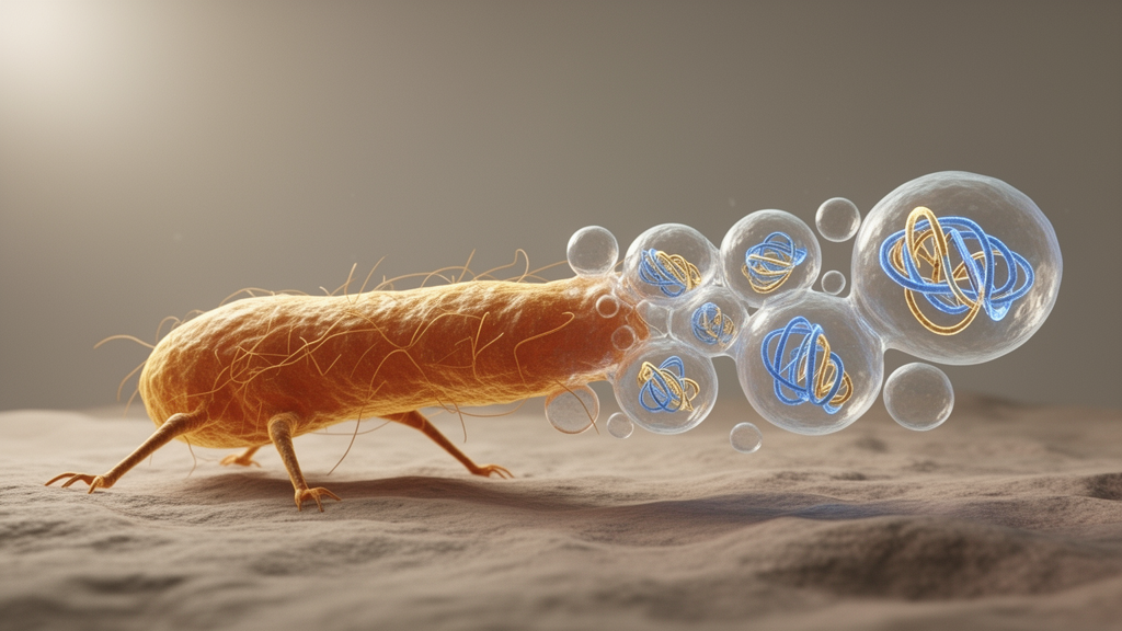
Cancer cells do not merely crawl through tissue; they leave cargo behind. A new study published in Nature Cell Biology describes a previously unrecognized class of large extracellular vesicles, dubbed motile vesicles or MoVes, that are shed by actively migrating cancer cells and carry a striking payload: the complete genomic DNA of the parent tumour cell, together with organelles and proteins tied to epithelial–mesenchymal transition, the cellular program that lets epithelial cancer cells turn invasive and motile. The findings, from Thupten Tsering of McGill University and colleagues across McGill and Weill Cornell Medicine, add a new branch to the growing family tree of extracellular vesicles and suggest that cell migration itself may double as a mechanism of molecular release.
The team first spotted the structures with scanning electron microscopy in the pericellular region of highly migratory breast cancer cell lines, including MDA-MB-231, 4T1 and MCF7. The vesicles measured roughly 2 to 5 micrometres across, placing them among the largest extracellular vesicles described. Notably, the non-tumorigenic breast epithelial cell line MCF10A produced none at all. When the researchers transformed MCF10A cells with the HRAS oncogene, creating the MCF-10AT line, cell motility rose from 0.24 to 0.809 micrometres per minute, a difference the authors report as statistically significant, and MoVes biogenesis appeared alongside it. That pairing of transformation, motility and vesicle shedding set the template for the rest of the study.
Live-cell imaging and electron microscopy traced how the vesicles form. MoVes arise from the trailing edge of mesenchymally migrating cells: a thin rear projection extends, a vesicle buds at its distal tip, remains tethered by filamentous connections, then detaches and is left behind on the substrate as the cell pulls away. The vesicles stayed anchored where they were deposited; the researchers observed no free-floating MoVes, suggesting their release is spatially restricted and possibly substrate-associated. Transmission electron microscopy of cross-sectioned vesicles revealed a crowded interior containing mitochondria, Golgi apparatus, endoplasmic reticulum, lysosomes and actin. Time-lapse imaging even hinted at chemoattractant-like behaviour, with neighbouring cells drawn toward the sites where MoVes were released, an activity absent in the less migratory MCF10A cells under identical conditions.
Generality mattered here, and the authors tested it broadly. Every cancer cell line examined produced MoVes, though with variable abundance: uveal melanoma lines 92.1 and MP41 and cervical CaSki cells emitted them more frequently than colorectal HT-29 and SW620 cells, while leiomyosarcoma lines showed low abundance. Cancer-associated fibroblasts shed more MoVes than normal fibroblasts. By contrast, MoVes were rarely observed in any non-cancerous cell type tested, including primary human normal choroidal melanocytes from three independent donors, immortalized human hepatocytes and intestinal epithelial cells. The authors are careful about the boundary: MoVes formation appears associated with cellular transformation and active migration rather than being strictly restricted to cancer cells, a nuance that matters for interpreting what these vesicles signify.
The genomic cargo is the headline result. Immunofluorescence staining showed MoVes enriched in double-stranded DNA, BrdU-incorporated replicative DNA and histone proteins, pointing to nuclear material inside. To probe DNA topology, the team treated small extracellular vesicles, large extracellular vesicles and MoVes with proteinase K and DNase I before lysis. DNase I reduced detectable DNA by roughly 98.8 percent in small vesicles and about 89 percent in large vesicles, but only about 65 percent in MoVes, indicating that a greater fraction of MoVes-associated DNA is protected from nuclease digestion, consistent with enclosure inside the vesicular lumen. Fragment analysis showed a prominent peak at 1,600 base pairs, and RNA profiling revealed ribosomal RNA peaks around 2 kilobases for the 18S subunit and around 5 kilobases for the 28S subunit, consistent with substantial incorporation of bulk cytoplasmic ribosomal content.
Whole-genome sequencing then confirmed the scope of that cargo. MoVes encapsulate the full genomic DNA of their parent 4T1 cells, including somatic mutations, with cancer-relevant mutations in Trp53, Pik3cg, Kit and Brca2 identified in both MoVes-derived DNA and matched cellular DNA. Mutations in the tumour suppressor Trp53 and the proto-oncogene Kit were validated independently by Sanger sequencing and droplet digital PCR. In practical terms, each vesicle recapitulates the mutational landscape of the tumour cell that released it, making MoVes non-lytic carriers of replicative, full-length genomic DNA. The authors note that DNA was detected both within and on the extracellular surface of the vesicles, and that a plausible origin is cytoplasmic double-stranded DNA, a byproduct of chromosomal instability and mitotic errors.
Proteomics sharpened the identity of the new vesicle class. Using differential centrifugation followed by ultracentrifugation, the researchers isolated three extracellular vesicle subtypes and compared them by liquid chromatography–tandem mass spectrometry, identifying 705 proteins at a 1 percent false discovery rate, with 427 shared across all subtypes. Among 46 proteins enriched in MoVes, FAM3C stood out. FAM3C, also known as ILEI, is a secreted interleukin-like protein with known epithelial–mesenchymal transition-inducing activity. Principal component analysis separated the vesicle subtypes cleanly and showed considerable molecular similarity between MoVes and blebbisomes, another large vesicle population, while immunofluorescence, western blotting and density gradient purification all confirmed FAM3C enrichment in MoVes. Gene ontology analysis placed MoVes proteins in redox homeostasis, oxidative stress response and lipid metabolism, with cellular components pointing to membrane, mitochondrion and endoplasmic reticulum.
Mechanistically, MoVes biogenesis proved dependent on migration and cytoskeletal remodelling. Pharmacological inhibitors of actin polymerization, the Arp2/3 complex, dynamin, Rho-ROCK signalling and non-muscle myosin II all cut migration speed by roughly 65 to 75 percent and markedly attenuated MoVes formation in breast cancer cells. Cytochalasin B primarily impaired early initiation, leaving cells stationary and rounded, whereas dynasore, Y-27632 and blebbistatin predominantly affected late initiation, producing elongated cells with defective rear-end detachment. The authors caution that these inhibitors can exert off-target effects and that their data do not establish whether dynamin is directly involved in vesicle emission. Notably, GW4869, an inhibitor of neutral sphingomyelinase 2 that blocks small vesicle biogenesis, did not affect MoVes emission, indicating a secretion mechanism distinct from classical exosome pathways. Knockdown of the myosin IIB gene Myh10, which reduces blebbisome release, did not significantly affect MoVes biogenesis, while Fam3c knockdown reduced both cell proliferation and MoVes emission.
The epithelial–mesenchymal transition connection ran in both directions. Blocking the TGFβ receptor with LY-364947 at a non-toxic concentration of 150 nanomolar inhibited TGFβ1-induced EMT, attenuated migration and produced complete suppression of MoVes biogenesis and MoVes-DNA release in 4T1 and MDA-MB-231 cells, while restoring epithelial markers such as E-cadherin and EpCAM. Conversely, inducing EMT in MCF10A cells with a commercial EMT-inducing medium supplement for 10 days produced a spindle-shaped morphology, a modest but statistically significant rise in migration speed from 0.27 to 0.42 micrometres per minute, and, importantly, the emission of MoVes from cells that normally produce none. Together these experiments support a model in which MoVes biogenesis is tightly linked to the EMT program and the migratory state it confers.
Functional and in vivo tests rounded out the picture, with caveats attached. MoVes carried mitochondria with intact membrane potential, and when applied to a mitochondrial DNA-depleted 4T1 ρ0 recipient line, MoVes, large vesicles and blebbisomes each increased basal and maximal respiration and mitochondria-dependent ATP production, with MoVes reaching statistical significance at P = 0.0228. In immunodeficient NSG mice, fluorescently labelled MoVes injected intravenously accumulated predominantly in the liver, like other vesicle subtypes, and were taken up by F4/80-positive Kupffer cells. But at higher doses, mice developed acute respiratory distress and died within minutes, with post-mortem imaging showing strong pulmonary accumulation, probably reflecting mechanical occlusion of lung microcapillaries roughly 1 to 2 micrometres in diameter. The authors list their limitations candidly: trypsin-based isolation may compromise surface protein composition, functional roles in vivo remain unexplored, large vesicle subtypes overlap considerably in size, and pharmacological inhibitors are not uniquely specific to MoVes. Even so, the study establishes MoVes as a distinct, migration-associated vesicle population capable of disseminating genomic DNA and organelles, and reframes cell migration as not only a means of movement but also a mechanism of molecular release and communication.
Subject of Research: A newly described population of large extracellular vesicles released by migrating cancer cells that carries whole genomic DNA and organelles
Article Title: Motile vesicles (MoVes) are a cancer cell migration-associated extracellular vesicle population
Article References: Tsering, T., Nadeau, A., Ghasemi, M., Mousovich-Neto, F., Tobias, G., Chen, Y., Kienzle, L., Dickinson, K., Douanne, N., Serganova, I., Zappasodi, R., Matei, I., Lyden, D., & Burnier, J. V. (2026). Motile vesicles (MoVes) are a cancer cell migration-associated extracellular vesicle population. Nature Cell Biology. https://doi.org/10.1038/s41556-026-02097-2
Image Credits: AI Generated
DOI: 10.1038/s41556-026-02097-2
Keywords: extracellular vesicles, MoVes, cancer cell migration, epithelial-mesenchymal transition, genomic DNA, whole-genome sequencing, mitochondrial transfer, FAM3C, proteomics, tumour biology, cell biology, Nature Cell Biology
News Source: Nathaniel Bowman. (October 28, 2026). Cancer cells on the move shed giant vesicles packed with whole genomes. Scienmag.



