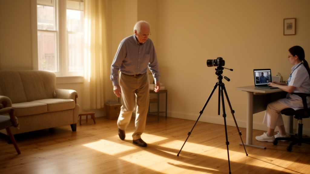
Neurology has always been a discipline of watching. Before a formal clinical encounter begins, a neurologist may notice a foot hesitating at a doorway, a stride shortening on the way to a chair, or a subtle flattening of facial animation during a greeting. Movement disorders specialists watch for amplitude, speed, asymmetry and overflow, in the clinic, in corridors, sometimes as early as the waiting room. Despite decades of progress in molecular imaging, genetics, fluid biomarkers and artificial intelligence, Parkinson’s disease is still most often first recognised through this language of movement: in what the body does, how it does it, and what it no longer quite does. Video has long been central to this practice, helping clinicians teach, document, compare and remember, and shaping the visual norms through which human variation is understood. It carries scientific value, but also moral weight, because it documents disease and a person living with it.
Artificial intelligence has now arrived at the core of this visual tradition. Computer vision tools powered by convolutional neural networks can document and measure movement from an ordinary video, including smartphone recordings, without expensive or complex hardware. Not long ago, this would have seemed an audacious prospect for a field so intimately linked to visual documentation. The promise is seductive: take what neurologists already do, carefully observe movement, and make it measurable, repeatable and available beyond clinic walls and the constraints of tight schedules. For Parkinson’s disease, a condition that fluctuates over hours and days and evolves over years, this is an attractive proposition. The disease is still assessed largely through brief, artificial clinical encounters. Clinicians ask patients how they have been over weeks or months, then score a few minutes of performance under fluorescent lights, often at a time of day that may not represent usual function, not to mention the well-known good day effect. Everyone knows this practice is born of a lack of better tools, and that it is insufficient.
The literature gives reason for optimism. Recent systematic reviews show that Parkinson’s disease is the most frequently studied movement disorder in computer vision research, that gait is the most common task analysed, and that diagnostic accuracies frequently exceed 80 percent. Open-source and commercial platforms suggest the field is moving from bespoke engineering demonstrations toward shared clinical tools. Several studies, many published in npj Parkinson’s Disease, have shown that computer vision may inform both clinical decision-making and the pathophysiological investigation of parkinsonian bradykinesia across body parts, therapy conditions and care settings. Beyond bradykinesia, computer vision has enabled video-based quantification of tremor amplitude and frequency, the pull test and facial expressivity, often with close agreement to reference methods such as motion capture and accelerometry. A camera in a clinic, home or aged-care facility, it seems, could soon reveal meaningful motor information without wires, markers or specialist hardware.
Yet the growing number of publications should not be confused with equivalent progress toward clinically valid digital biomarkers. Showing that an algorithm can distinguish patients from controls, reproduce a clinical score or perform well within a selected dataset is an important first step, but it is not sufficient. A video-derived measure intended for clinical use must undergo the same scrutiny as any other biomarker or clinical outcome measure. It should demonstrate test-retest reliability, acceptable measurement error, construct validity, responsiveness to clinically meaningful change, and robustness across populations, recording environments and disease stages. Crucially, researchers need to understand whether a change in the output reflects a true change in the patient rather than variation in camera position, lighting, task execution or model performance. Without this deeper clinimetric work, technical accuracy can easily be mistaken for biological or clinical validity. This may explain why the field has produced many models and feasibility demonstrations, but comparatively few, if any, clinically mature digital biomarkers.
The potential, for all the fascination with these advances, is clear: better measurement, earlier detection, remote care, trial enrichment and more equitable access to specialist assessment. But realising that potential depends on addressing a pressing challenge: the translation of technical performance into valid measurement, clinical action and patient benefit. Beyond practical issues such as who requests, analyses and interprets video outputs, harder questions follow. A 10 percent change in finger-tapping amplitude means little until we know what it means for someone’s daily life. Increasing sensitivity may also shift the boundaries of what we call prodromal or pre-symptomatic disease, raising questions about disclosure, follow-up, responsibility and reimbursement. Patients must be involved in deciding how they feel about being filmed for diagnosis or monitoring. And any tangible benefit must be legible to both patients and their physicians, not just to a model. These are the implementation questions that separate a research instrument from a clinical tool, and addressing them will require implementation science from the outset, including evaluation of acceptability, feasibility, workflow fit, equity, sustainability and unintended consequences across diverse care settings.
A tool may be technically accurate and still be clinically irrelevant. It may correlate with a rating scale but fail to change a decision. It may add measurement without adding meaning. With the broad availability of wearable technology, medicine is generating a growing number of digital outcomes, yet despite their technical validity, adoption in clinical studies and guidelines remains very limited. For computer vision to avoid this fate, it must be built around defined clinical decisions and care pathways rather than computational possibility, with explainability taking centre stage. Explainability should be treated not as a cosmetic layer added after model development but as a clinical requirement tested with its intended users. Parkinson’s care does not need more numbers in the abstract. It needs help with practical, complex questions: Is levodopa still helping? Is gait changing because of Parkinson’s disease, arthritis, cognition, pain or environment? Is this treatment effect large enough to matter to the person living with the disease?
There are harder questions still. If a computer vision algorithm detects deterioration before the patient does, but no effective intervention or care response is available, have we actually helped? Do patients even want this? If a model flags early Parkinson’s disease in someone not yet diagnosed, who carries responsibility for that knowledge? The ethical risks extend beyond the misuse of video data. They include the danger that patients become increasingly visible to health systems without becoming meaningfully better supported by them. Surveillance without response is not care. If a system can detect falls risk, wearing-off, freezing or deterioration, it must be connected to a pathway for timely action. Without such a pathway, computer vision risks becoming a sophisticated way of exposing disease-related need while leaving that need unmet.
This shifts the scientific goalposts away from technical benchmarks, bespoke coding-intensive solutions and clinically meaningless error metrics, and toward deployable, user-friendly and interpretable tools that generate outcomes of value to both doctors and patients. The value of such digital biomarkers extends beyond technical performance: it emerges from rigorous testing against the decisions and outcomes they are intended to inform. This aligns with the broader digital biomarker literature in Parkinson’s disease, which emphasises that digital measures require evidence of reliability, validity, interpretability, regulatory readiness and integration into care pathways before they can become clinically useful. More speculatively, computer vision can also inspire clinicians to reconsider how they visually assess patients. If computer vision can quantify a clinical gesture such as finger tapping with this much nuance, should that precision not be leveraged to measure real-world function directly, moving toward ecologically valid neuroscience? Or what if a machine’s attention could teach our own, by pointing clinicians’ eyes to the regions of a video frame it found most informative for a given clinical outcome? In both scenarios, explainability is paramount. Clinicians are more likely to trust and act on outputs that resemble clinical reasoning than on black-box abstraction: a report visualising reduced amplitude, progressive decrement and increasing irregularity during finger tapping is more interpretable, and likely more acceptable, than an opaque probability derived from a model clinicians struggle to understand, let alone explain to a patient. Such explanation must also be faithful to how the model reached its conclusion, rather than merely plausible visualisations generated after the event. The goal is not to replace the neurologist’s eye but to extend it, to slow it down, render it more quantitative and objective, and let it attend to moments it might have missed.
Future directions should therefore be ambitious but not naive. Researchers can imagine movement vital signs: brief standardised videos captured at home or before clinic, summarising gait speed, turning, arm swing or facial expressivity. They can imagine remote specialist augmentation, in which rural clinicians, aged-care staff or general neurologists capture structured video and receive an interpretable, clinically governed movement summary, not a diagnosis from a machine, but a better map for human care. Other fields that have already travelled this path offer important lessons. Radiology did not become digital simply because images could be processed; it built picture archiving systems, reporting standards, accreditation, quality assurance and clinical governance. Diabetes care did not integrate continuous glucose monitoring merely because glucose could be measured continuously; it became useful when patients, clinicians, devices, reimbursement systems and therapeutic algorithms converged around action. Aviation and other high-stakes contexts emphasise that automation becomes safe when embedded in systems that account for human behaviour, fatigue, error, accountability and trust. In this vein, the movement disorders field now needs its own infrastructure for visual, measurement-based medicine, including not only technical standards but implementation frameworks capable of identifying where, for whom and under what conditions computer vision adds value.
The next phase of computer vision in Parkinson’s disease should be defined not by the number of movements that can be extracted from video, but by the number of video-derived measures that withstand rigorous biomarker validation and improve a specified clinical decision. That demands three things: consensus standards for video acquisition, metadata, reporting and validation; prospective implementation studies testing whether computer vision changes clinical decisions, outcomes or patient experience, with outputs co-designed with clinicians, patients and carers; and governance encompassing bias and failure-mode audits, regulatory and reimbursement pathways matched to real use cases, and explicit responsibility when systems fail. Publication standards should rise accordingly: retrospective performance in a curated dataset should be considered an entry point, not an endpoint. Still, the most important future may be quieter than all of this. Could computer vision help patients feel believed? Many people with Parkinson’s describe changes that are intermittent, subtle or hard to name. They know when walking has become subjectively less automatic, when buttoning a shirt feels harder, or when medication subtly starts to wear off. A sensitive visual tool could make these experiences visible, not replacing the patient’s narrative but standing alongside it. The measure of computer vision will not be merely its tracking accuracy, but whether it leaves the person in front of us better cared for, and perhaps better believed. Now comes the harder work: making computer vision part of medicine, not merely part of research. Just as Parkinson’s disease alters movement in fractions, with a little less amplitude and a little more effort, computer vision allows us to measure those fractions earlier, more sensitively and more faithfully. Its future will be determined not by how precisely algorithms quantify movement, but by whether those measurements improve decisions, transform outcomes, and ultimately help people with Parkinson’s disease live better lives.
Subject of Research: Computer vision applications for measuring and clinically translating Parkinson's disease motor assessment from video
Article Title: Computer vision, Parkinson’s disease, and the unfinished work of clinical translation
Article References: Alty, J., & Friedrich, M. U. (2026). Computer vision, Parkinson’s disease, and the unfinished work of clinical translation. npj Parkinson's Disease, 12(1), Article 253. https://doi.org/10.1038/s41531-026-01577-4
Image Credits: AI Generated
DOI: 10.1038/s41531-026-01577-4
Keywords: Computer, vision, Parkinson, disease, unfinished, work, clinical, translation, scientific research
News Source: Cassandra Pierce. (October 18, 2026). Cameras Can See Parkinson’s Movement, But Turning That Into Care Is Harder. Scienmag.