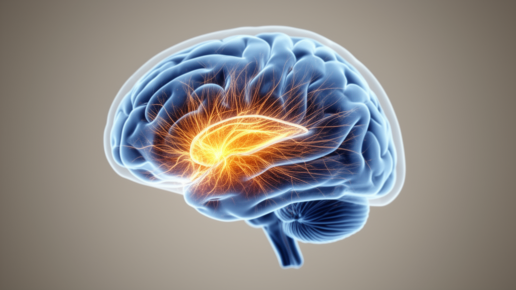
Chronic pain is increasingly understood not as the simple aftermath of persistent injury signals but as a disorder of distributed brain networks. At the center of that reorganization, according to a new editorial published in BioPsychoSocial Medicine, sits the medial prefrontal cortex (mPFC), a region that links the sensory experience of pain with its affective, cognitive, and stress-related dimensions. The authors, Marco Cascella, Mario Montedoro, and Alessandro Vittori, argue that the mPFC functions as a system-level integrative hub, but they also deliver a sobering caveat: neither mPFC-centered models nor artificial intelligence approaches are ready for the clinic.
The mechanistic case for the mPFC rests on converging lines of evidence. Functional magnetic resonance imaging studies in people who underwent transcutaneous electrical stimulation to induce secondary hyperalgesia found that higher mPFC activation was associated with smaller hyperalgesic fields, hinting at a protective, top-down modulatory role. Intriguingly, mPFC activity was also inversely related to pharmacological responsiveness: individuals showing reduced descending pain modulation, presumably reflecting impaired mPFC-mediated control, responded better to systemic lidocaine. This positions mPFC activity as a candidate neurobiological correlate of analgesic responsiveness, though the editorial insists it should never be treated as an isolated, validated clinical biomarker.
Beneath the imaging signals lies a rich neurochemical landscape. Magnetic resonance spectroscopy studies have linked chronic pain to reduced glutamate levels in the mPFC, changes that align with elevated anxiety, depression, and avoidance behaviors. Dopaminergic modulation adds another layer: inputs from the ventral tegmental area to the mPFC regulate cortical plasticity, aversive learning, and motivational states, and in neuropathic pain models, phasic activation of these mesocortical projections reduces mechanical hypersensitivity. At the circuit level, dopaminergic signaling enhances mPFC neurons projecting to the ventrolateral periaqueductal gray, a key node of the descending pain control system.
The microcircuitry tells an equally detailed story. Fast-spiking parvalbumin-positive interneurons, which coordinate prefrontal network oscillations and top-down control, show region- and layer-specific maladaptive plasticity in chronic pain states. Experimental models indicate that enhanced feed-forward inhibition from these interneurons can suppress mPFC pyramidal neurons projecting to the periaqueductal gray, weakening descending inhibitory control, while loss of inhibitory input in other mPFC subregions drives pyramidal hyperexcitability and affective dysregulation. Adding to this, µ-opioid receptors located on GABAergic interneurons, rather than on glutamatergic pyramidal neurons, appear to mediate stress-induced analgesia by disinhibiting mPFC neurons that project to the periaqueductal gray.
Functional neuroimaging supplies the network-scale view. Resting-state fMRI studies have identified convergent alterations in amplitude of low-frequency fluctuations, regional homogeneity, and large-scale functional connectivity. Longitudinal neuroimaging has revealed a profound reorganization during the transition from acute to chronic pain: a relative attenuation of activity in primary sensory cortices accompanied by increased engagement of prefrontal-limbic regions, including the mPFC and amygdala, with preferential involvement of default mode network nodes such as the mPFC and precuneus. Strikingly, these shifts occur even without measurable changes in pain intensity, underscoring the dissociation between subjective pain ratings and large-scale network dynamics, a finding with major implications for how pain should be modeled.
The reciprocal connectivity between the mPFC and amygdala forms a crucial regulatory loop. The mPFC exerts inhibitory control over amygdala output through excitatory projections to intercalated cells, while the basolateral amygdala sends glutamatergic projections back to the mPFC, creating a negative feedback circuit. In chronic pain models, hyperactive amygdala output dampens mPFC activity, impairing cognitive control over nociception and enabling amplification of negative affect, a maladaptive cycle that partially overlaps with circuit-level features of post-traumatic stress disorder and major depression, though the authors caution this reflects shared network vulnerabilities rather than interchangeable pathophysiology. In cancer-related chronic pain and other severe syndromes, functional disintegration between the mPFC and limbic structures has been associated with diminished top-down regulation.
Stress physiology adds yet another dimension. The mPFC influences the hypothalamic-pituitary-adrenal axis and the autonomic nervous system, modulating the termination, rather than the peak, of glucocorticoid release, aided by dense glucocorticoid receptor expression. In chronic migraine, altered mPFC-hypothalamus connectivity within the default mode network correlates with subjective pain intensity, potentially reflecting a loss of cognitive reappraisal capacity.
It is against this backdrop that artificial intelligence enters the picture. The editorial frames multimodal AI integration, combining machine learning and deep learning, as a potential way to decode these multifaceted neurobiological signatures, enabling automated, objective pain assessment and adaptive therapy monitoring in research settings. Feature-level, model-level, and decision-level fusion strategies can combine neuroimaging, neurophysiological signals, behavioral measures, and clinical variables, while architectures such as convolutional neural networks, recurrent networks, and graph-based models extract hierarchical representations of brain activity. Generative AI approaches, agent-based models, and digital twins may further support hypothesis generation by simulating individualized network dynamics and exploring counterfactual perturbations of mPFC-centered circuits.
Yet the authors are emphatic that these technologies are enabling and exploratory methods, not near-term clinical solutions. AI output remains dependent on incompletely defined phenotypes, indirect surrogate labels, and limited external validation. Methodological obstacles include bias and confounding from cohort composition, limited generalizability across clinical populations and acquisition settings, and the risk of data leakage in multimodal pipelines. Defining a valid ground truth for pain remains intrinsically problematic because pain is multidimensional, subjective, and context-dependent. Robust clinical models would require multi-center data collection, protocol harmonization, external validation across independent cohorts, longitudinal analyses, and systematic evaluation of interpretability and failure modes, steps that are still largely lacking.
Generalizability poses further boundary conditions. Most mPFC-centered models derive from adult populations, and their extension to children, older adults, patients with psychiatric comorbidities, cancer-related pain, cognitive impairment, or those under chronic pharmacological treatment cannot be assumed. Emerging evidence from large-scale developmental neuroimaging suggests altered brain signatures, including trends toward reduced mPFC activity and disrupted connectivity, may precede multisite pain in children, but pediatric pain remains embedded in ongoing prefrontal maturation and is currently supported by limited empirical evidence. The editorial concludes that mPFC-centered and AI-enabled approaches should be interpreted as conceptual tools for hypothesis generation and refining pain models, not as instruments for immediate clinical translation, and that bridging the gap to practice will require rigorously designed, longitudinal, and developmentally informed studies alongside transparent validation frameworks.
Subject of Research: The role of the medial prefrontal cortex in chronic pain network dysfunction and the exploratory use of artificial intelligence in pain research.
Article Title: The medial prefrontal cortex as an integrative hub in chronic pain: network mechanisms and the enabling role of artificial intelligence
Article References: Cascella, M., Montedoro, M., & Vittori, A. (2026). The medial prefrontal cortex as an integrative hub in chronic pain: network mechanisms and the enabling role of artificial intelligence. BioPsychoSocial Medicine, 20(1), Article 6. https://doi.org/10.1186/s13030-026-00357-z
Image Credits: AI Generated
DOI: 10.1186/s13030-026-00357-z
Keywords: chronic pain, medial prefrontal cortex, artificial intelligence, neuroimaging, default mode network, multimodal integration, pain modulation, digital twins, GABAergic interneurons, HPA axis, medial, prefrontal
News Source: Cassandra Pierce. (October 30, 2026). Brain’s Pain Hub: Why the Medial Prefrontal Cortex Resists Quick Clinical Answers. Scienmag.