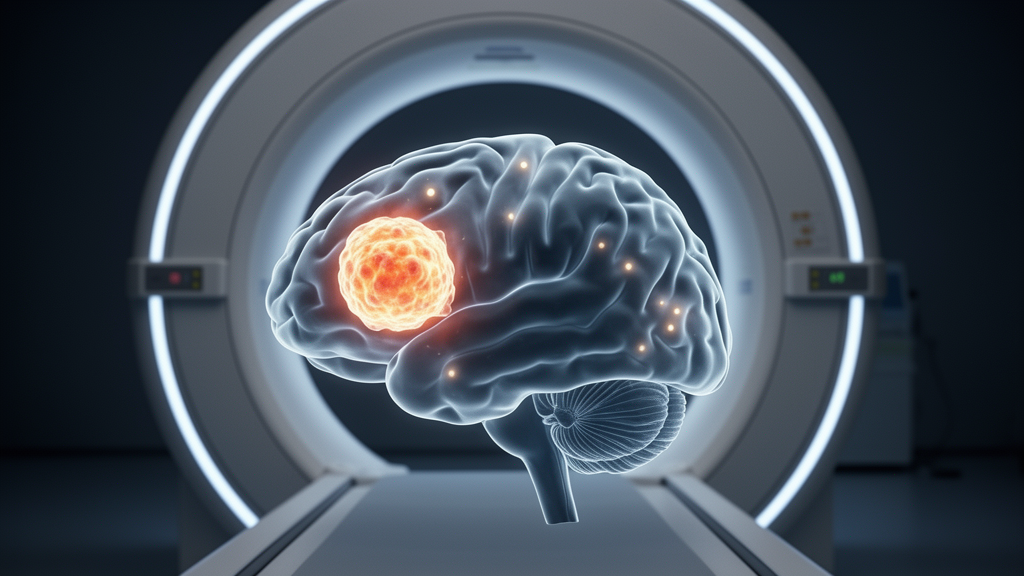
For decades, the standard playbook for treating aggressive brain tumors has rested on a single assumption: that the boundaries of the disease can be seen clearly on a magnetic resonance imaging scan. A new prospective pilot study from a team at Sunnybrook Health Sciences Centre and the University of Toronto now provides some of the strongest North American evidence yet that this assumption is wrong in a substantial fraction of patients. In the FETSMaRT trial, published in the Journal of Neuro-Oncology, researchers enrolled thirty patients with World Health Organization grade 3 or grade 4 high-grade gliomas and compared the tumor volumes that radiation oncologists would normally draw on MRI with the volumes revealed by a combined PET/MRI scan using a radioactive amino acid tracer. The results show a systematic and statistically significant underestimation of metabolically active tumor by conventional MRI, a finding with direct consequences for how radiotherapy targets are defined.
The tracer at the heart of the study is O-(2-[18F]fluoroethyl)-L-tyrosine, commonly abbreviated as FET. Unlike the gadolinium-based contrast agents used in routine MRI, which highlight regions where the blood-brain barrier has broken down, FET is taken up by tumor cells through amino acid transporters that are upregulated in glioma tissue. This means the PET signal reflects metabolic activity rather than vascular leakage, allowing clinicians to visualize infiltrating tumor cells that extend beyond the enhancing core seen on contrast-enhanced T1-weighted MRI. International guidelines from the European Association of Nuclear Medicine, the European Association of Neuro-Oncology and the RANO group have already endorsed amino acid PET for glioma imaging, but prospective data from North American centers integrating FET-PET directly into radiotherapy planning have remained scarce. FETSMaRT was designed to fill that gap.
The study design was deliberately rigorous for a pilot. Thirty patients, all scheduled to receive post-operative radiotherapy, underwent a combined FET-PET/MRI acquisition before starting treatment. Crucially, the radiation oncologist who delineated the standard MRI-based target volumes was blinded to the PET data. Only afterward did the team generate a second set of targets derived from the FET-PET/MRI information, enabling a head-to-head, volumetric comparison of gross tumor volumes, clinical target volumes and planning target volumes. The median age of the cohort was sixty years, seventy-seven percent had glioblastoma, the most aggressive grade 4 form, and sixty percent carried a methylated MGMT promoter, a molecular marker associated with better chemotherapy response. This composition mirrors the typical high-grade glioma population seen in major treatment centers.
The headline numbers are striking. Discordance between the MRI-based and PET-derived gross tumor volumes occurred in seventeen of thirty patients, or 56.7 percent, while discordance at the clinical target volume level affected fifteen of thirty patients, exactly half of the cohort. Across the entire group, the FET-PET/MRI-derived volumes were consistently and significantly larger than the MRI-based ones. The gross tumor volume averaged 43.6 cubic centimeters on PET compared with 38.6 cubic centimeters on MRI, a difference that reached a p-value below 0.0001. The clinical target volume averaged 119.2 versus 110.8 cubic centimeters, with a p-value of 0.0045, and the planning target volume averaged 171.1 versus 159.6 cubic centimeters, with a p-value of 0.0003. In plain terms, the metabolic footprint of these tumors was reliably bigger than what the anatomical images suggested.
That consistency matters because of how radiotherapy for glioblastoma is actually delivered. Under current practice, shaped radiation beams are aimed at a target volume that expands outward from the visible tumor to account for anticipated microscopic infiltration, typically reaching a dose of sixty gray in thirty fractions over six weeks alongside the chemotherapy drug temozolomide. This treatment framework, established by the landmark Stupp trials of 2005 and 2009, has changed little in its imaging foundation even as radiation delivery technology has become dramatically more precise. If the biological target is systematically drawn too small, the most advanced linear accelerators in the world will still miss tumor cells that were never contoured in the first place.
The patterns of treatment failure recorded in the study reinforce that concern. Among patients whose disease progressed, the failures were predominantly central, meaning the tumor recurred within or near the high-dose radiation field in 60 percent of cases. Central failure is the classic signature of tumor cells that received radiation but survived, and it suggests that even the treated volume may not have covered the full metabolically active extent of disease. Historical dosimetric studies going back to the late 1990s have documented similar central recurrence patterns after three-dimensional conformal radiotherapy, and the FETSMaRT data add a metabolic imaging dimension to that long-standing problem.
The researchers also examined whether the size of the volume discrepancy predicted how quickly patients progressed. In the glioblastoma subgroup of twenty-three patients, Kaplan-Meier and Cox proportional hazards analyses related the magnitude of gross tumor volume and clinical target volume discordance to time to progression. The direction of the association was consistent: patients with larger discordance values tended to progress sooner. However, in this small pilot cohort neither association reached statistical significance. The authors are careful on this point, describing the findings as hypothesis-generating exploratory data that justify a larger prospective trial rather than proof of a survival effect. Small sample size is the obvious limitation, and the study was funded by a grant from the Brain Tumor Foundation Canada precisely to build the case for that definitive trial, which is registered as NCT06499831.
The broader context makes the timing of this work significant. A multicentre randomized European trial known as GLIAA, reported in The Lancet Oncology, recently compared FET-PET-guided re-irradiation against contrast-enhanced MRI guidance in patients with recurrent glioblastoma, adding momentum to the idea that amino acid PET should guide radiation decisions. Meanwhile, a separate Canadian phase 2 trial called UNITED has explored MRI-guided adaptive radiotherapy for high-grade glioma, and the TROG 18.06 FIG study in Australia has been prospectively evaluating FET imaging in glioblastoma across multiple centers. Taken together, these efforts signal a field-wide reassessment of the anatomical imaging paradigm that has governed brain tumor radiotherapy for a generation, with metabolic imaging positioned as the likely successor.
What would change for patients if FET-PET/MRI were adopted into routine planning? The volumetric differences documented in FETSMaRT, on the order of five to twelve cubic centimeters depending on the target level, are large enough to alter dose distribution in clinically meaningful ways, particularly for tumors located near eloquent brain regions where expanding the target risks harming functional tissue. Hybrid PET/MRI scanners, which acquire both modalities in a single session with perfectly registered images, remove much of the logistical friction that historically made PET integration cumbersome. The study authors note that deidentified participant data and analysis code will be available on reasonable request, supporting independent verification and pooled analyses as the evidence base grows.
For the roughly 77 percent of the cohort facing glioblastoma, a disease with a median survival that has crept only modestly above the fifteen-month mark despite two decades of intensifying therapy, the promise of a more accurate target is not an incremental technicality. Every cubic centimeter of metabolically active tumor that escapes the radiation field is a potential seed of recurrence, and central failures remain the dominant pattern of relapse. FETSMaRT does not prove that PET-guided contours will extend survival, but it establishes with prospective, blinded, quantitative rigor that conventional MRI systematically underestimates the biological extent of high-grade glioma in more than half of patients. The stage is now set for the definitive randomized trial that the Toronto team argues their pilot data demand, one that could finally align the physics of modern radiotherapy with the biology of the tumors it is meant to eradicate.
Subject of Research: Comparison of 18F-FET-PET/MRI and MRI-based radiotherapy target volumes in high-grade glioma
Article Title: Volumetric discordance between ¹⁸F-FET-PET/MRI and MRI-based radiotherapy target volumes in high-grade glioma: a prospective pilot study (FETSMaRT)
Article References: Volumetric discordance between ¹⁸F-FET-PET/MRI and MRI-based radiotherapy target volumes in high-grade glioma: a prospective pilot study (FETSMaRT). (n.d.). https://doi.org/10.1007/s11060-026-05831-w
Image Credits: AI Generated
DOI: 10.1007/s11060-026-05831-w
Keywords: 18F-FET-PET, glioblastoma, high-grade glioma, radiotherapy planning, PET/MRI, target volume delineation, tumor recurrence, neuro-oncology, medical imaging, clinical trial, MGMT methylation, radiation oncology
News Source: Nathaniel Bowman. (October 15, 2026). Brain Scans Miss Hidden Tumor Spread, Landmark Glioma Imaging Trial Reveals. Scienmag.