A new short communication in EJNMMI Physics, published on 15 October 2026, reports a controlled way to measure that distortion and to judge how well existing correction methods fix it.
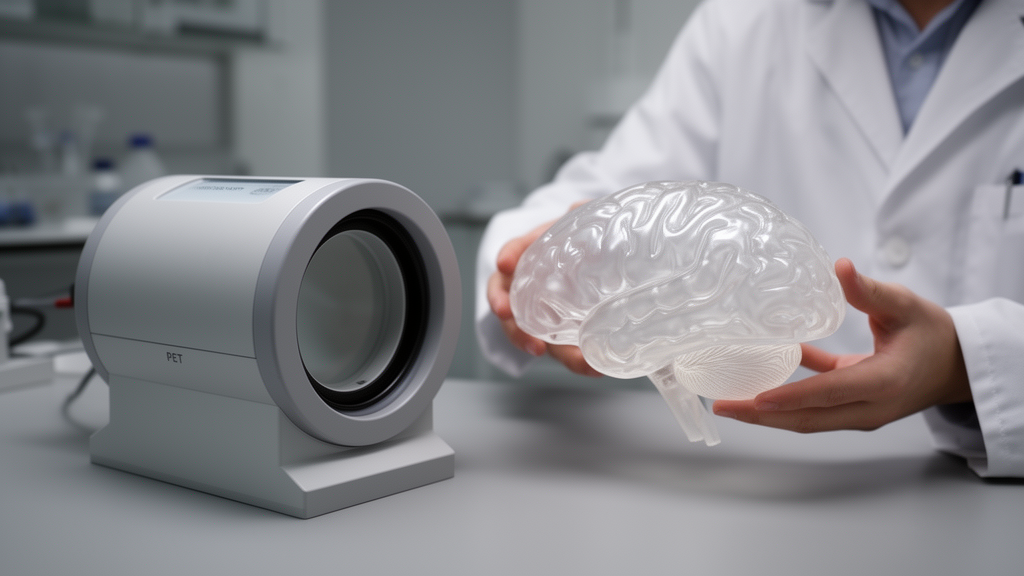
The research team, led by Maria Agnese Pirozzi and Mario Quarantelli with colleagues including Eugenio Di Giorgio, Anna Prinster, Luigi Mansi, Bruno Alfano and Marco Spadafora, used an anatomical brain phantom called StepBrain (SB).
The phantom was scanned to simulate clinical amyloid PET studies across a range of gray-to-white matter ratios (GWR) from 0 to 120 percent. This range was designed to reproduce the clinical spectrum of amyloid uptake patterns, from amyloid-negative to strongly positive scans, giving the researchers a known ground truth against which measurement error could be calculated.
From the simulated scans, the team derived two widely used quantitative measures: the gray-to-white matter ratio and the Standardized Uptake Value Ratio (SUVR), normalized to the cerebellum. Both were computed from an atlas-based volume-of-interest set, first on uncorrected images and then after applying partial volume effect correction (PVEc).
Four correction approaches were compared: a two-compartment method (2C), a three-compartment method (3C), a modified three-compartment method (m3C), and the geometric transfer matrix (GTM) technique.
The results were striking. Uncorrected GWR values showed a mean absolute inaccuracy of 14.1 percent, and uncorrected SUVR values were off by 19.8 percent on average. After applying the correction methods, these errors fell to as low as 6.1 percent for GWR and 7.6 percent for SUVR.
Accuracy and precision differed significantly among the four correction methods for both outcome measures, with linear mixed models analysis yielding F statistics above 95 in all comparisons. The modified three-compartment method and the geometric transfer matrix delivered the highest accuracy and precision of the approaches tested.
According to the authors, the 3C-, m3C- and GTM-based corrections significantly improved tracer ratio accuracy without a relevant loss of precision, supporting their use in the quantitative analysis of clinical amyloid PET studies. Because the StepBrain phantom reproduced realistic amyloid PET distributions while providing an exact ground truth, it offers a practical validation platform for correction algorithms before they are applied to patient data.
As a phantom-based simulation study, the work does not itself establish clinical performance in patients, but it provides a rigorous benchmark for the methods that underpin amyloid burden measurement in both research and clinical PET practice.
Subject of Research: Validation of partial volume effect correction methods for amyloid PET using an anatomical brain phantom.
Article Title: Application of an anatomical brain phantom in the evaluation of partial volume effect correction of amyloid PET studies
Article References: Pirozzi, M. A., Di Giorgio, E., Prinster, A., Mansi, L., Alfano, B., Spadafora, M., & Quarantelli, M. (2026). Application of an anatomical brain phantom in the evaluation of partial volume effect correction of amyloid PET studies. EJNMMI Physics. https://doi.org/10.1186/s40658-026-00956-3
Image Credits: AI Generated
DOI: 10.1186/s40658-026-00956-3
Keywords: amyloid PET, partial volume effect, brain phantom, SUVR, gray-to-white matter ratio, GTM, nuclear medicine, quantitative imaging, Alzheimer's research, image correction, Application, anatomical
News Source: Cassandra Pierce. (October 28, 2026). Brain Phantom Study Puts Amyloid PET Correction Methods to the Test. Scienmag.