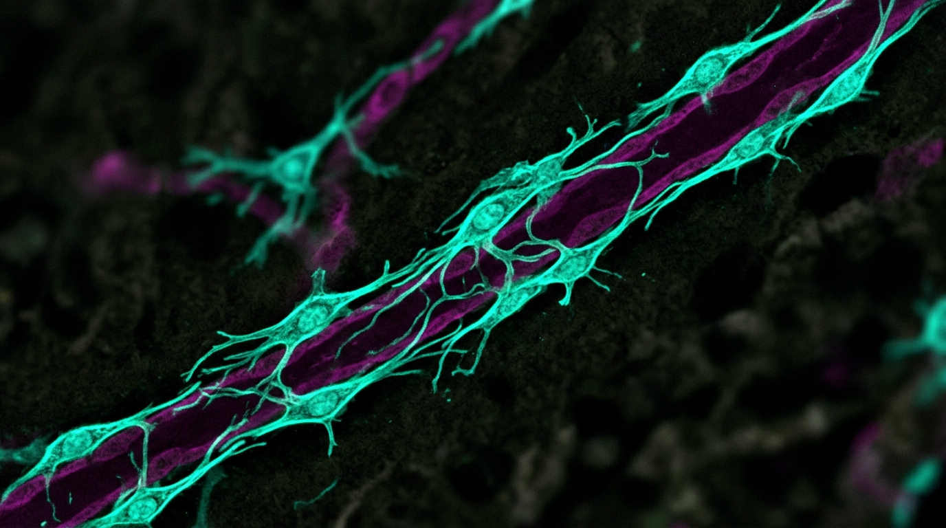
Pericytes, the mural cells wrapped around the smallest blood vessels, have long been treated as a single class of vascular support cell. A new comparative study in Frontiers in Cell and Developmental Biology argues that this picture is incomplete. By isolating pericytes from two very different organs of the mouse, the brain and the lung, and running both populations through the same isolation and flow cytometry pipeline, researchers found that the two cell populations share a conserved mesenchymal-associated core yet differ sharply in the organization of their canonical pericyte markers, with CD90 emerging as the principal feature separating brain-derived from lung-derived cells.
The team, led by Isabel María Martínez, Antonio Martínez-Sanchez, Miguel Blanquer and Rut Valdor at the University of Murcia and the Biomedical Research Institute of Murcia, focused on CD146, an adhesion receptor long used to identify perivascular cells. Seminal work had previously linked CD146-positive perivascular cells to mesenchymal stromal progenitors in multiple adult tissues, and CD146 has been associated with enhanced mesenchymal and regenerative potential. In the brain, the receptor also coordinates endothelial cell-pericyte interactions required for blood-brain barrier formation and maturation. Whether CD146-defined pericyte subsets from organs with different developmental origins and physiological demands carry equivalent mesenchymal characteristics, however, had not been systematically tested.
The experimental design was deliberately symmetrical. Pericytes were obtained from the microvasculature of mouse brains, excluding the cerebellum, brainstem, meninges and olfactory bulb, and from whole lungs of pools of four to six male and female C57BL/6 mice aged six to eight weeks. After mechanical dissociation and enzymatic digestion with collagenase A and DNase I, the microvessel-enriched fractions were cultured to passage five, then enriched for CD146-positive cells using magnetic-activated cell sorting with CD146 MicroBeads. Both the retained CD146-positive fraction and the CD146-negative counterpart were then immunophenotyped by multiparametric flow cytometry at the sixth passage, using two marker panels that covered CD146, the canonical pericyte markers PDGFRβ, RGS5, NG2 and α-SMA, and the mesenchymal-associated markers CD90, CD44 and CD105. A third panel confirmed the absence of endothelial and hematopoietic contamination by checking CD31, CD34 and CD45.
The results showed that CD146 cleanly separates two non-endothelial pericyte populations in both organs, but the two fractions are not equivalent. In brain-derived pericytes, roughly 83 percent of CD146-positive cells co-expressed PDGFRβ and RGS5, and about 52 percent were double-positive for PDGFRβ and α-SMA, while NG2 co-expression was comparatively low at around 24 percent. The CD146-negative brain fraction was markedly weaker across the board: about 36 percent co-expressed PDGFRβ and RGS5, roughly 14 percent PDGFRβ and α-SMA, and only about 6 percent PDGFRβ and NG2. Quantitative analysis confirmed that the proportion of positive cells for all evaluated canonical markers, and their mean fluorescence intensity, was significantly higher in the CD146-positive population than in the CD146-negative counterpart.
Lung-derived pericytes followed the same broad pattern with different numbers. In the CD146-positive pulmonary fraction, approximately 73 percent of cells co-expressed PDGFRβ and RGS5, but co-expression with α-SMA and NG2 was much lower, at about 19 percent and 6 percent respectively. The CD146-negative pulmonary fraction again showed reduced canonical marker expression, with around 49 percent of cells co-expressing PDGFRβ and RGS5, about 14 percent PDGFRβ and α-SMA, and roughly 5 percent PDGFRβ and NG2. Notably, NG2 expression remained low in the lung and did not differ significantly between CD146-positive and CD146-negative subsets, unlike the brain. When the CD146-positive fractions from both tissues were analyzed in parallel, brain-derived pericytes displayed significantly higher proportions of RGS5-positive, α-SMA-positive and NG2-positive cells than lung-derived pericytes, despite similarly high PDGFRβ levels in both, revealing a tissue-specific phenotypic organization of the pericyte compartment.
The mesenchymal side of the comparison produced the study’s most striking finding. Mesenchymal stromal cells are conventionally characterized by markers such as CD90, CD105 and CD44, and pericytes have been reported to share key phenotypic features with them. In both organs, CD146-positive pericytes showed significantly higher expression of mesenchymal-associated markers than their CD146-negative counterparts. In the brain, approximately 60 percent of both the PDGFRβ-positive and RGS5-positive subsets within the CD146-positive fraction co-expressed CD90, whereas the CD146-negative fraction showed a markedly reduced proportion of these double-positive cells. CD105 and CD44, by contrast, remained consistently high across all evaluated brain subsets. In the lung, the majority of CD146-positive pericytes co-expressed CD105 and CD44 together with either PDGFRβ or RGS5, and about 40 percent of both PDGFRβ-positive and RGS5-positive subsets also co-expressed CD90. Quantitative analysis confirmed that all three mesenchymal markers were significantly enriched within the CD146-positive pulmonary fraction.
Direct comparison between tissues sharpened the picture. CD44 expression was similarly high in brain- and lung-derived CD146-positive pericytes, marking it as a conserved mesenchymal-associated feature regardless of tissue origin. CD105 expression was largely comparable, with only a modest but significant reduction in the PDGFRβ-positive pulmonary subset. CD90, however, was consistently and significantly reduced in both PDGFRβ-positive and RGS5-positive pulmonary pericytes compared with their brain-derived counterparts, making it the principal marker distinguishing the two populations. The authors note that CD90 is not merely a phenotypic label; it has been implicated in the regulation of stemness, proliferation and lineage commitment in mesenchymal progenitors, and in mechanosensing and cell-matrix adhesion. They hypothesize that the lower CD90 level in pulmonary pericytes might reflect an adjustment to the distinct biomechanical properties of the lung microenvironment, while stressing that their descriptive data cannot establish functional consequences and that future studies will be needed to test this idea.
To test whether the mesenchymal phenotype survives increasingly stringent definitions of pericyte identity, the researchers used sequential gating to isolate a triple-positive population co-expressing PDGFRβ, α-SMA and RGS5 within the CD146-positive fractions. Both brain- and lung-derived cells in this gate showed robust, near-ubiquitous CD44 and CD105 expression, and the tissue-specific difference in CD90 was maintained. When the analysis was restricted further to cells co-expressing all four pericyte-associated markers examined, CD146, PDGFRβ, RGS5 and α-SMA, the proportion of CD90-positive cells rose above the double-positive levels, reaching approximately 80 percent in brain-derived pericytes and 65 percent in lung-derived pericytes. Both brain- and lung-derived CD146-defined populations showed negligible expression of the endothelial and hematopoietic markers CD31, CD34 and CD45, confirming their non-endothelial, non-hematopoietic perivascular stromal identity.
The authors are careful about what these findings do and do not establish. Because freshly isolated pericytes yield too few cells for the extensive and repeated phenotypic analyses required, the comparison was performed on primary pericytes expanded ex vivo to passage five or six. They acknowledge that culture conditions and the loss of the native vascular microenvironment can influence pericyte phenotypic fidelity: transcriptomic work cited in the paper shows that cultured mouse brain pericytes at passage five form a more homogeneous population whose profile resembles embryonic more than adult pericytes. The differences identified should therefore be read as tissue-of-origin-associated phenotypic properties retained under standardized ex vivo conditions, not as a complete representation of the native in vivo state. The isolation approach also integrates pericytes from different anatomical and microvascular regions within each organ, so region-specific heterogeneity within either organ is not resolved. The authors suggest that immunofluorescence, RNA in situ hybridization and spatially resolved molecular profiling, including human biopsy specimens when available, would provide complementary validation while preserving the native perivascular niche.
Even with those caveats, the study offers a framework with implications beyond basic vascular biology. Pericytes are active regulators of inflammatory and remodeling responses in both organs: pulmonary pericytes participate in leukocyte recruitment, cytokine signaling, vascular remodeling and fibrotic responses in chronic inflammatory lung disease, pulmonary hypertension and acute lung injury, while brain pericytes regulate blood-brain barrier integrity, neuroinflammation and tissue repair after injury. The marked difference in α-SMA expression between the two tissues, readily detected in a substantial proportion of brain pericytes but markedly lower in pulmonary populations, may reflect both tissue-specific mural cell identity and distinct states of pericyte plasticity or activation, since α-SMA can also be upregulated during pericyte activation and phenotypic switching in vascular remodeling and inflammation. The authors also connect their findings to chaperone-mediated autophagy, a mechanism their laboratory has linked to the maintenance of pericyte homeostatic and mesenchymal-associated phenotypes, and to observations that pericytes at different distances from glioblastoma lesions display distinct immune-regulatory properties. By defining a conserved mesenchymal-associated core with tissue-specific organization, the work establishes a comparative baseline for investigating how tissue-specific pericyte states contribute to tissue homeostasis, inflammatory disease and tumor-associated microenvironments.
Subject of Research: Tissue-specific marker and mesenchymal signatures of murine brain and lung pericytes
Article Title: Comparative characterization of lung and brain pericytes reveals tissue-specific marker and mesenchymal signatures
Article References: Martínez, I. M., Martínez-Sanchez, A., Blanquer, M., & Valdor, R. (2026). Comparative characterization of lung and brain pericytes reveals tissue-specific marker and mesenchymal signatures. Frontiers in Cell and Developmental Biology, 14, Article 1950106. https://doi.org/10.3389/fcell.2026.1950106
Image Credits: AI Generated
DOI: 10.3389/fcell.2026.1950106
Keywords: pericytes, CD146, mesenchymal stromal cells, brain, lung, flow cytometry, CD90, RGS5, PDGFRβ, alpha-SMA, pericyte heterogeneity, murine study
News Source: Cassandra Pierce. (November 6, 2026). Brain and Lung Pericytes Share a Mesenchymal Core but Diverge in Tissue-Specific Markers. Scienmag.