In the realm of biomedical imaging, fluorescence and luminescence technologies have emerged as groundbreaking tools for non-invasive visualization at the cellular and tissue levels. However, the inherent challenges posed by tissue photon scattering, optical absorption, and autofluorescence often cloud the clarity of these images, causing significant background noise and spectral overlap. To transcend these limitations, scientists have shifted their focus toward the second near-infrared (NIR-II) window that spans wavelengths from 1000 to 2000 nm, a spectral region recognized for its remarkable capacity to mitigate scattering and absorption effects in biological tissues. This property enables higher resolution and deeper penetration in imaging applications.
Crucially, an optimal excitation and emission pairing within this NIR-II region around 808 nm excitation and emissions exceeding 1500 nm has attracted intense research interest. The rationale hinges on the employment of commercially available 808 nm lasers that contribute to efficient excitation, combined with emissions in the NIR-IIb subwindow (>1500 nm), which further reduce tissue scattering and minimize water-induced heating. The utilization of lanthanide-doped nanomaterials, especially those based on erbium ions (Er³⁺), is highly coveted as their intrinsic emission near 1530 nm fits almost perfectly within this subwindow, granting them exceptional potential for deep-tissue imaging.
Yet, one of the fundamental roadblocks hindering the advancement of lanthanide-based NIR-II probes is the inherently limited absorption cross-section of Er³⁺ ions at the excitation wavelength around 808 nm, a consequence of f-f forbidden electronic transitions. This drastically reduces the efficiency of direct excitation and luminescence generation, making it challenging to develop probes with sufficiently bright emissions for practical imaging applications. To overcome this intrinsic limitation, one promising strategy involves employing organic dye molecules featuring absorption cross-sections orders of magnitude greater than those of lanthanide ions. These dyes can act as antennae, absorbing excitation light efficiently and transferring energy to the lanthanide ions in a process termed dye sensitization.
Despite the promise of dye sensitization, the efficiency of energy transfer between the dye donors and lanthanide acceptors is tethered to the delicate balance dictated by Förster resonance energy transfer (FRET) principles. FRET efficiency sharply declines with increasing donor-acceptor distance, confining the effective energy transfer to nanometer proximities and, consequently, limiting the amount of lanthanide ions effectively excited when the dye is bound to the nanoparticle surface. This spatial constraint has long posed a formidable challenge: how to maximize energy transfer throughout the entirety of a densely doped lanthanide core from surface-bound organic dyes.
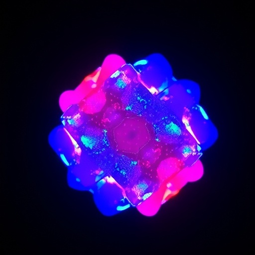
Addressing this fundamental challenge, the research team led by Professor Yulei Chang at the State Key Laboratory of Luminescence Science and Technology, Changchun Institute of Optics, Fine Mechanics and Physics, Chinese Academy of Sciences, has pioneered an ingenious shell engineering approach. Their innovative design harnesses the NaErF₄ core, rich in Er³⁺ ions, and couples it with a strategically engineered shell containing a high concentration of Yb³⁺ ions acting as mediating energy relays. This 808 nm-excited system intends to optimize the core’s absorption while balancing energy transfer and surface quenching effects, resulting in significantly amplified luminescent output at around 1525 nm.
The core-shell nanostructure features a NaErF₄ core enveloped by a NaYF₄ shell doped with 50% Yb³⁺ ions, subsequently conjugated with the organic dye indocyanine green (ICG). This architecture facilitates a cascaded energy transfer mechanism that diverges from the conventional direct dye-to-Er³⁺ energy pathway. Instead, the cascade incorporates the sensitizing agent ICG transferring energy to Yb³⁺ ions in the shell, which then relay this energy to the Er³⁺ ions in the core. This multi-step relay effectively collects and chaperones the excitation energy, minimizing nonradiative losses and invigorating the luminescence of the lanthanide ions with unparalleled efficiency.
Paramount to this system’s success is the doping concentration of Yb³⁺ in the shell. The 50% doping level strikes an optimal equilibrium: it sufficiently amplifies energy transfer between the dye and lanthanide core while simultaneously mitigating deleterious surface quenching phenomena that plague heavily doped assemblies. This delicate balancing act results in a near 2000-fold enhancement of luminescence intensity at 1525 nm compared to traditional inert-shell counterparts, signaling a paradigm shift in the engineering of lanthanide-based probes.
To unravel the photophysical underpinnings of their design, the researchers meticulously examined the excited-state lifetimes under 980 nm emission across different nanoparticle configurations with and without conjugated ICG dye. The lifetimes for Er@Y@ICG (where the shell contains only Y³⁺ ions), Y@50Yb@ICG (shell containing 50% Yb³⁺, but no Er³⁺ in the core), and Er@50Yb@ICG were systematically compared. Intriguingly, Er@Y@ICG displayed the shortest emission lifetime, indicative of rapid nonradiative decay and limited energy transfer. In contrast, Y@50Yb@ICG showed markedly prolonged lifetimes owing to efficient dye-Yb³⁺ energy transfer without Er³⁺ involvement. Most compellingly, Er@50Yb@ICG’s intermediate but appreciably longer lifetime underscores the efficacy of the Yb³⁺-mediated cascade transfer, amassing kinetic evidence for the hypothesized ICG → Yb³⁺ → Er³⁺ energy relay process.
The energy transfer from ICG to the nanoparticles is remarkably efficient, with lifetime measurements revealing a dramatic decrease in ICG’s excited-state lifetime from 883 ps to 84 ps upon conjugation with the Er@50Yb construct, translating to a staggering approximately 90% energy transfer efficiency. Further studies employing cyclooctatetraene (COT) as a triplet-state quencher clarified that energy transfer predominantly occurs from the singlet excited state of the ICG dye. This pathway cleverly sidesteps complications arising from triplet-state losses, which can otherwise attenuate the emission efficacy in many photoluminescent systems.
Additionally, the 50% Yb³⁺ doping in the shell addresses and suppresses the concentration quenching that typically hinders luminescence in Er³⁺-rich cores, significantly enhancing the population of the Er³⁺ 4I13/2 energy level. This state is directly responsible for the characteristic 1525 nm emission corresponding to the Er³⁺ 4I13/2 → 4I15/2 transition. The result is an unprecedented near 2000-fold increase in emission brightness relative to bare core structures, exceeding the performance benchmarks of conventional Nd³⁺-sensitized lanthanide systems.
Following successful fabrication, the nanoprobes were PEGylated to enhance biocompatibility and circulation stability for in vivo applications. Deploying these probes for vascular imaging in murine models yielded pioneering results in the NIR-IIb window. Images achieved a spatial resolution marked by a full width at half maximum of 218 μm and exhibited an excellent signal-to-background ratio of 3.09, facilitating the visualization of vascular structures with remarkable clarity and contrast. Moreover, the nanoprobes demonstrated a favorable blood circulation half-life of 53 minutes, aligning well with the demands of dynamic vascular imaging.
This trailblazing dye-sensitized cascaded energy transfer methodology presented by Professor Chang and colleagues has not only bridged one of the major gaps in lanthanide nanoparticle design but also established a powerful platform for future development of deep-tissue NIR-IIb imaging probes. By pioneering an effective relay mechanism enhanced via structural and compositional optimization, this work paves the way toward next-generation nanoprobes capable of exceptional luminescence performance with broad biomedical implications. The profound implications for vascular research and potentially for enhanced diagnostic imaging are poised to accelerate breakthroughs in clinical and fundamental science.
The implications of this research extend beyond fluorescence brightness enhancement. The fundamental insights into nanoarchitecture-mediated energy dynamics may very well inspire novel designs across luminescent materials, photovoltaic devices, and photocatalysis, where efficient light harvesting and transfer are paramount. The interplay of organic and inorganic components, balanced doping, and multi-step cascade energy transfer exemplifies the sophistication achievable in nanophotonics.
With continued innovation leveraging such principles, future nanoprobes may offer unprecedented imaging depth, resolution, and functional specificity while maintaining biocompatibility and safety. This research marks a defining milestone not only in the pursuit of brighter, deeper, and more selective imaging probes but also in the broader quest to harness photonic phenomena at the nanoscale for transformative applications in science and medicine.
Subject of Research: Development of dye-sensitized lanthanide nanoparticles for enhanced near-infrared luminescence aimed at deep tissue imaging.
Article Title: Dye-sensitized cascaded energy transfer for amplified 1525 nm luminescence in highly doped lanthanide nanoparticles
Web References: 10.1038/s41377-026-02302-9
Image Credits: Fei Long et al.
Keywords
Near-infrared imaging, lanthanide nanoparticles, dye sensitization, energy transfer cascade, Erbium ions, Ytterbium doping, fluorescence lifetime, NIR-IIb window, organic dyes, indocyanine green, PEGylation, deep tissue luminescence, biomedical imaging, nanoprobe design.
Tags: 1525 nm luminescence enhancement808 nm laser excitation in bioimagingcascaded energy transfer in nanomaterialsdeep tissue near-infrared imagingdye-sensitized energy transfer mechanismserbium ion emission propertiesfluorescence background noise reductionlanthanide-doped nanoparticles for biomedical imagingNIR-II window fluorescence imagingNIR-IIb subwindow applicationsnon-invasive cellular imaging techniquesovercoming tissue scattering and absorption

