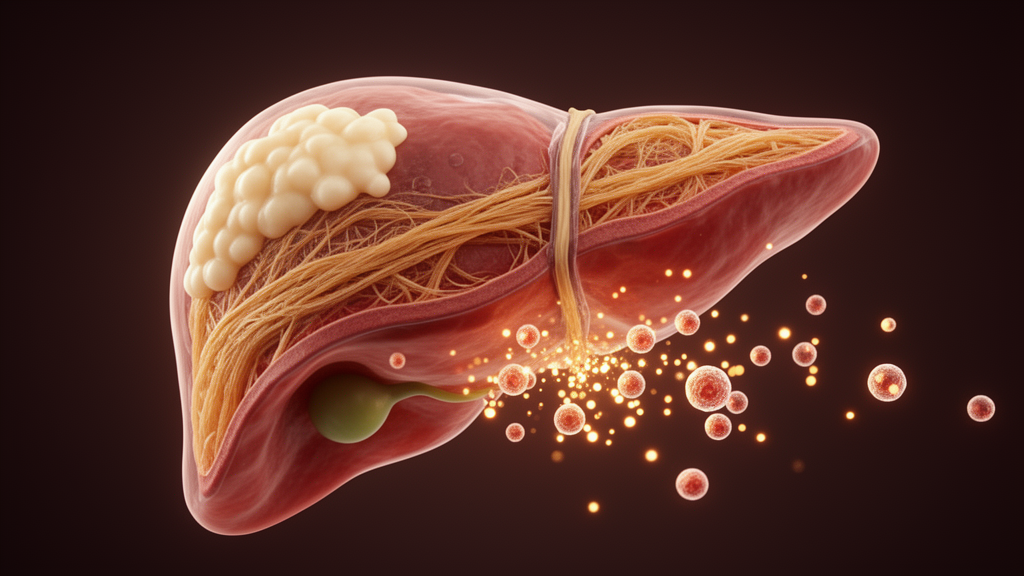
Researchers have identified and independently validated a set of eleven proteins carried by tiny membrane-bound particles circulating in the blood that tracks with advanced fibrosis in metabolic dysfunction-associated steatohepatitis, or MASH. The work, published in Cell & Bioscience as an open-access article, suggests that the protein cargo of circulating extracellular vesicles may capture liver-derived signals that conventional blood tests miss.
Advanced fibrosis is a major determinant of liver-related mortality in MASH, the aggressive form of fatty liver disease linked to metabolic dysfunction. Yet the non-invasive tests currently used in clinical practice lack specificity for staging fibrosis and offer limited resolution of the disease biology occurring in the liver itself. The study set out to test whether proteomic analysis of plasma extracellular vesicles could yield a validated biomarker signature for advanced fibrosis while also shedding light on underlying mechanisms.
The team analyzed plasma extracellular vesicles from 222 adult participants in the NIDDK NASH Clinical Research Network, all with biopsy-confirmed MASLD. Participants had a mean age of 52 years and a mean body mass index of 33 kg/m2, and they represented the extremes of the histologic fibrosis spectrum: individuals with no fibrosis, staged F0, and individuals with advanced fibrosis, staged F3 to F4. A discovery cohort of 112 participants, matched for age, sex and body mass index, was used to identify the initial signature, which was then tested in an independent, unmatched validation cohort of 110 participants.
Proteomic profiling was performed using unbiased high-resolution mass spectrometry with data-independent acquisition and parallel accumulation serial fragmentation, known as DIA-PASEF. The researchers then carried out differential abundance analysis and developed machine-learning classifiers using extracellular vesicle proteins alone or in combination with clinical variables.
The analysis produced an eleven-protein extracellular vesicle signature associated with advanced fibrosis. Among the proteins were QSOX1, FCGR3A, PIGR and TGFBI, whose abundance patterns tracked with fibrosis severity across the sampled stages, a pattern consistent with fibrosis-associated disease biology. The authors note that these abundance relationships were observed in the fibrosis stages sampled, which spanned the extremes of the spectrum rather than every intermediate stage.
A key finding concerned where the signals come from. The extracellular vesicle cargo showed significantly greater enrichment for liver-associated signals compared with bulk plasma or serum proteomic datasets.
The diagnostic performance was tested in the validation cohort. An Elastic Net model integrating extracellular vesicle proteins with clinical variables achieved an area under the receiver operating characteristic curve of 0.807, with a 95 percent confidence interval of 0.722 to 0.893. The corresponding clinical model, built without the vesicle proteins, reached an AUROC of 0.645 in the same cohort. These figures describe discrimination between the F0 and F3-F4 groups in this specific cohort and are model estimates rather than established clinical performance.
The authors conclude that circulating extracellular vesicle proteomics identified and independently validated a signature associated with advanced fibrosis in MASH and captured liver-enriched signals that were less apparent in bulk plasma or serum analyses. They position the approach as complementary to existing fibrosis assessment methods and as a potential source of biologically informative signals associated with liver disease, rather than as a replacement for current tools.
Several caveats frame the findings. The study compared the extremes of the fibrosis spectrum, so performance in intermediate stages, in broader populations, or against existing non-invasive tests head-to-head remains to be established. The article was also shared early to provide faster access to peer-reviewed, accepted research; this version is citable and carries a permanent DOI but is subject to further edits and will be replaced by the final Version of Record.
By showing that this compartment carries measurably stronger liver-associated protein signals than unsorted blood proteomics, and that a machine-learning model built on those signals improves discrimination of advanced fibrosis beyond clinical variables alone, the work outlines a route toward blood-based fibrosis staging grounded more directly in the biology of the injured liver.
Subject of Research: Extracellular vesicle proteomics as a biomarker approach for advanced fibrosis in metabolic dysfunction-associated steatohepatitis
Article Title: Circulating extracellular vesicle proteomics identifies and validates a signature of advanced fibrosis in MASH
Article References: Pirrotte, P., Garcia–Mansfield, K., Yates, K. P., Wu, X., Lovell, B., Piras, I. S., Agrawal, A., Martinez, M. N., Sharma, R., & DiStefano, J. K. (2026). Circulating extracellular vesicle proteomics identifies and validates a signature of advanced fibrosis in MASH. Cell & Bioscience. https://doi.org/10.1186/s13578-026-01660-6
Image Credits: AI Generated
DOI: 10.1186/s13578-026-01660-6
Keywords: extracellular vesicles, proteomics, MASH, liver fibrosis, biomarkers, mass spectrometry, NASH Clinical Research Network, machine learning, Elastic Net, MASLD, non-invasive testing, QSOX1
News Source: Drew Townsend. (October 29, 2026). Blood vesicle proteins flag advanced scarring in fatty liver disease. Scienmag.