Cell therapies may stop or reverse the pain and disability of degenerative disc disease and the loss of material between vertebrae, according to Duke University scientists.
The health conditions affect thousands of Americans. To use cell therapies, however, scientists have to keep the cells alive, synthesize the appropriate replacement material and get it to the right place in a patient’s spine. With newly made biomaterials from Duke’s Pratt School of Engineering, that goal could be closer.
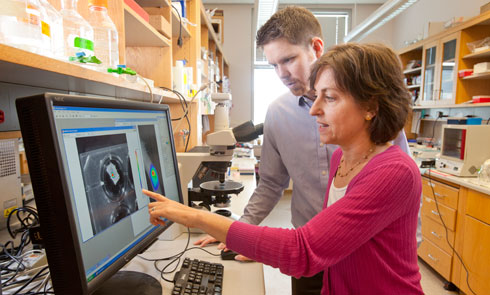
In a proof-of-concept study published online in the journal Biomaterials, graduate student Aubrey Francisco and biomedical engineering professor Lori Setton describe a new biomaterial designed to deliver a booster shot of reparative cells to the nucleus pulposus, or NP — the jelly-like cushion naturally found between spinal discs. The NP tissue distributes pressure and provides spine mobility, helping to relieve back pain.
“Our primary goal was to create a material that would be liquid at the start, gel after injection in the disc space and keep the cells in the location where they’re needed,” Setton said. “Our second goal was to create a material that would provide the delivered cells with the environmental cues to promote their persistence and biosynthesis.”
Disc degeneration is a common problem as people age. Over time, the soft, compressible discs that work as the spine’s shock absorbers break down. Although this intervertebral disc degeneration can occur anywhere along the spine, it mainly happens near the neck and lower back, causing intense pain. Individuals with this condition can also develop herniated discs, osteoarthritis or spinal narrowing, known as spinal stenosis.
Previous lab research has shown that re-implanting NP cells, or even stem cells, can delay disc degeneration. Several companies already offer cell delivery strategies, but the methods are poor and ineffective. “They allow the cells to quickly migrate out of and away from the injection site,” Francisco said.
The Duke team’s delivery strategy keeps the cells in place and provides cues that mimic laminin, a protein in native nucleus pulposus tissue. Laminin is normally found in juvenile but not degenerated discs and allows injected cells to attach and remain in place with the delivered biomaterial. Laminin may also enable the cells to survive longer and produce more of the appropriate extracellular matrix or structural underpinning of the discs that help stop degeneration, Setton said.
The researchers, funded by the National Institutes of Health, developed a gel mix designed to reintroduce NP cells to the intervertebral disc (IVD) site. The gel mixes together three components: the protein laminin-111 that has been chemically modified and two polyethylene glycol (PEG) hydrogels that can attach to the modified laminin. Separately, these substances remain in a liquid state. The gel, however, holds the cells in place upon injection.
The researchers worked with postdoctoral scholar Robby Bowles to tag NP cells with the bioluminescent marker luciferase and track their location. They then injected the gel into rats’ tails just as a surgeon would deliver cells to a patient. After puncturing the tail’s thin outer layer, they held the needle in place for one minute, delivered the injection to the rat’s IVD site, and closed the injection spot. The solution began to solidify after five minutes and was completely set at 20 minutes.
Images of the luciferase biomarker showed that more than 14 days after injection significantly more cells remained in place when delivered within the biomaterial carrier compared to cells delivered in a liquid suspension. Francisco explained that using the currently available cellular delivery strategies, 100 percent of the injected NP cells leak out of an IVD site within three to four days after injection.
The results, although preliminary, could have a positive impact on the future of cell therapy, Setton said. More work, however, is still needed to optimize equipment and models that deliver cells to a larger IVD sites closer to human size, the researchers said.
“The concept is that these cells will be promoted to produce matrix that can support tissue regeneration or arrest degeneration,” Setton said. “Additional studies that evaluate disc height or matrix hydration following cell delivery would be important to achieve this goal. There’s definitely interest and certainly real potential there.”
Collaborators from Duke University Medical Center were William Richardson, M.D., a spine surgeon in the Department of Orthopaedic Surgery, and Farshid Guilak, professor of orthopaedic surgery and biomedical engineering. Robert Mancino, a 2013 bachelor’s degree recipient from the Pratt School of Engineering, worked on this study as part of his independent research.
Story Source:
The above story is reprinted from materials provided by Duke University, Whitney Howell.

