A simple ultrasound probe pressed against the temple may reveal more about an acute stroke than clinicians previously appreciated. In a new study from Dr. Hasan Sadikin General Hospital in Bandung, Indonesia, researchers found that patients whose transcranial Doppler scans showed narrowing of the middle cerebral artery arrived at the hospital with substantially more severe neurological deficits than patients whose scans looked normal. The finding, published in Artery Research, offers a potentially valuable tool for settings where advanced brain imaging is not immediately available, while stopping well short of claiming that the artery abnormality itself causes worse strokes.
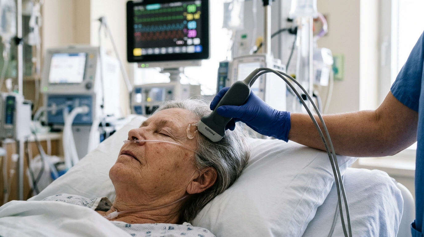
The study examined 50 consecutive adults with acute ischemic stroke who underwent transcranial Doppler examination of the middle cerebral artery between January and June 2023. Transcranial Doppler, or TCD, is a non-invasive bedside technique that measures blood flow velocity and waveform characteristics within intracranial arteries, primarily the mean flow velocity and peak systolic velocity along the M1 segment of the middle cerebral artery at depths of 45 to 65 millimeters. Because it requires only an acoustic window at the temple rather than a large scanner, it can be deployed quickly in emergency settings, though its accuracy depends on operator skill and adequate acoustic windows.
The results were striking in their consistency. TCD-detected middle cerebral artery stenosis was identified in 34 of the 50 patients, or 68 percent, while 16 patients, or 32 percent, showed no such abnormality. When the researchers compared admission scores on the National Institutes of Health Stroke Scale, the standard clinical measure of neurological impairment, the difference between the groups was pronounced. Patients with detected stenosis had a median NIHSS score of 10, with an interquartile range of 8 to 14, compared with a median of 4.5 among patients without stenosis, with an interquartile range of 3.75 to 5. The difference was statistically significant, with a p-value below 0.001.
Expressed as continuous averages, the gap persisted. The mean NIHSS score was 10.47 plus or minus 3.45 in the stenosis group versus 4.56 plus or minus 1.41 in the non-stenosis group, a difference of roughly 5.5 points on a scale where each point represents measurable neurological impairment. In categorical analysis, stenosis was more frequently observed among patients with moderate-to-severe strokes, while patients without detected stenosis were more often found in the mild category. Across the whole cohort, nine patients, or 18 percent, had mild strokes, 40 patients, or 80 percent, had moderate strokes, and one patient, or 2 percent, had a severe stroke by the study’s predefined thresholds.
What makes the finding notable is what the two groups did not differ on. Age was statistically indistinguishable between patients with and without detected stenosis, with means of 58.26 plus or minus 11.26 years and 56.44 plus or minus 9.92 years respectively. Hypertension, present in 79.4 percent of the stenosis group and 93.8 percent of the non-stenosis group, diabetes mellitus, dyslipidemia, smoking, previous stroke history, and cardiac disease all failed to show significant differences between the groups. The one demographic exception was sex: male patients were more frequently represented in the stenosis group, at 58.8 percent versus 25.0 percent, a difference the authors flag as suggestive but not proof that male sex independently predicts the artery abnormality, given the small sample and absence of multivariable adjustment.
The TCD criteria used to classify stenosis were hemodynamic rather than directly visual. A normal middle cerebral artery typically shows mean flow velocities between 30 and 80 centimeters per second. Stenosis of 50 percent or more was defined by mean flow velocity above 100 to 120 centimeters per second or peak systolic velocity above 140 to 180 centimeters per second. Severe or hemodynamically significant stenosis required mean flow velocity exceeding 140 centimeters per second, or an affected-to-contralateral velocity ratio greater than 2.0. Supporting features included flow turbulence visible as spectral broadening, reduced pulsatility index distal to the narrowing, or sudden loss of the velocity signal at a site of occlusion.
Crucially, the authors emphasize that a TCD-detected abnormality is not synonymous with intracranial atherosclerotic disease. Elevated flow velocity and abnormal spectral patterns can arise in several vascular conditions, and TCD alone cannot determine the underlying vascular etiology. The stenosis finding was therefore treated strictly as a hemodynamic observation. This distinction matters because the clinical consequences of altered flow depend on factors TCD does not directly measure, including collateral circulation, infarct location and size, systemic blood pressure, and the brain’s capacity for autoregulatory compensation.
The anatomical relationship between the detected abnormality and the index infarction adds another layer of complexity. Among the 34 patients with detected stenosis, 58 percent had ipsilateral lesions and 42 percent had bilateral lesions. A contralateral or anatomically unrelated abnormality may not directly explain deficits caused by an infarction in a different vascular territory, so analyses distinguishing ipsilateral from contralateral findings are important for interpreting clinical relevance. The authors note this interpretation remains a hypothesis because collateral status, infarct volume, and cerebral perfusion were not systematically evaluated in the study.
Several limitations constrain how far the conclusion can travel. The retrospective cross-sectional design prevents causal inference, and the single-center sample of 50 patients limits statistical power and generalizability. TCD-detected stenosis was not systematically confirmed with computed tomography angiography, magnetic resonance angiography, or digital subtraction angiography, so misclassification cannot be excluded. The analysis was unadjusted and did not account for infarct volume, infarct territory, collateral circulation, extracranial carotid disease, systemic hemodynamic conditions, previous antithrombotic treatment, or reperfusion treatment. The study also evaluated association with admission severity rather than functional outcome or longitudinal deterioration, so the findings should not be read as evidence of prognostic value.
Within those constraints, the study supports TCD’s potential role as a complementary bedside assessment of intracranial hemodynamics in acute ischemic stroke, particularly where advanced vascular imaging is not immediately available. The authors suggest that TCD findings should be interpreted alongside clinical examination and structural or vascular neuroimaging when available, and they call for larger prospective multicenter studies combining TCD with CTA or MRA, infarct characterization, collateral assessment, and longitudinal outcomes to determine whether TCD-detected abnormalities provide independent information beyond established clinical and imaging predictors. For now, the message is measured: an abnormal bedside Doppler signal in the middle cerebral artery may flag patients presenting with greater neurological impairment, but it is a clue to be weighed with other evidence, not a verdict on its own.
Subject of Research: Association between transcranial Doppler-detected middle cerebral artery stenosis and admission stroke severity in acute ischemic stroke
Article Title: Association Between Transcranial Doppler–Detected Middle Cerebral Artery Stenosis and Stroke Severity in Acute Ischemic Stroke
Article References: Amalia, L., & Andaru, A. S. (2026). Association Between Transcranial Doppler–Detected Middle Cerebral Artery Stenosis and Stroke Severity in Acute Ischemic Stroke. Artery Research, 32(1), Article 33. https://doi.org/10.1007/s44200-026-00134-x
Image Credits: AI Generated
DOI: 10.1007/s44200-026-00134-x
Keywords: transcranial Doppler, middle cerebral artery, acute ischemic stroke, NIHSS, stroke severity, intracranial stenosis, cerebral hemodynamics, blood flow velocity, bedside ultrasound, Indonesia, cross-sectional study, vascular imaging
News Source: Ophelia Keating. (November 6, 2026). Bedside Doppler Scan Flags Worse Strokes: Artery Finding Tied to Higher Severity Scores. Scienmag.