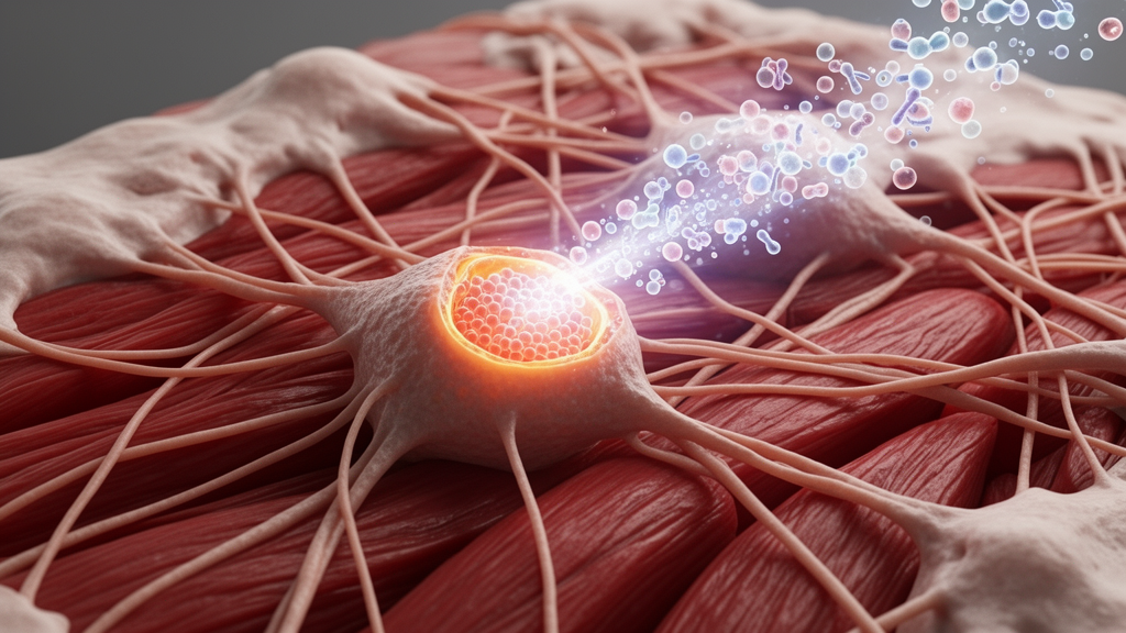
Cardiac fibrosis has long been viewed as one of the most stubborn problems in cardiovascular medicine, largely because the cells responsible for it are remarkably resilient once they switch into their scar-producing state. Fibroblasts are the most abundant cell type in the heart after muscle cells, and when injury occurs—whether from a heart attack, chronic high blood pressure, diabetes, or inflammatory conditions—they respond by proliferating and depositing extracellular matrix proteins such as collagen. In the short term this response is protective, forming a patch that keeps damaged tissue from rupturing. Over time, however, persistent fibroblast activation leads to excessive matrix deposition that stiffens the ventricular walls, impairs diastolic filling, disrupts electrical conduction, and limits oxygen delivery to working muscle. Epidemiological studies have consistently linked the degree of fibrosis to worse outcomes in heart failure with preserved ejection fraction, a condition that affects millions of people worldwide and for which few effective treatments exist. The absence of any approved drug that directly reverses or halts the scarring process itself has made the search for antifibrotic mechanisms a central priority in cardiovascular research.
The Stanford-led study adds an important dimension to this effort by focusing not on the fibroblasts themselves but on the signaling molecules that instruct them to activate. Adenosine is an attractive candidate for such scrutiny because its extracellular levels rise dramatically during ischemia, hypoxia, and tissue injury, conditions that coincide with the onset of fibrotic remodeling. Under normal circumstances adenosine serves as a metabolic distress signal, helping to restore blood flow and limit damage through vasodilation and anti-inflammatory effects. Yet the same molecule can also promote tissue remodeling in certain contexts, and the four known adenosine receptor subtypes—A1, A2A, A2B, and A3—mediate effects that vary widely depending on the cell type involved and the physiological setting. This pleiotropy has complicated pharmacological attempts to exploit the adenosine system, because a drug that blocks one subtype may produce unintended consequences in organs where that receptor performs beneficial functions.
What makes the new findings particularly notable is the demonstration that three of these subtypes, despite their conventional differences, funnel into a shared intracellular route. G protein-coupled receptors are traditionally understood to transmit signals through their associated G-alpha subunits, with each subtype coupling to a characteristic alpha protein that triggers distinct second-messenger cascades. The A1 receptor, for example, typically couples to Gi proteins that inhibit adenylate cyclase and lower intracellular cyclic AMP, while the A2A and A2B receptors couple to Gs proteins that raise cyclic AMP levels. Because these canonical pathways can produce opposing physiological outcomes, researchers have generally assumed that targeting them individually would yield subtype-specific effects. The discovery that all three subtypes also release G-beta-gamma subunits that converge on a common pro-fibrotic program in cardiac fibroblasts challenges that assumption and offers a mechanistic explanation for why single-subtype antagonists have shown inconsistent results in prior studies of fibrosis.
The G-beta-gamma complex has historically been treated as a passive partner in GPCR signaling, released from the G-alpha subunit upon receptor activation but regarded mainly as a modulator of other pathways rather than a primary driver of cellular responses. Over the past two decades, however, accumulating evidence has shown that G-beta-gamma can directly engage a variety of effectors, including phospholipase C isoforms, PI3 kinase, ion channels, and components of the MAP kinase cascade. In the context of cardiac fibroblasts, the new study links G-beta-gamma to downstream signals that govern fibroblast activation and the expression of scar-producing genes, positioning this complex as a nodal point where multiple upstream receptors meet. The demonstration that selectively inhibiting G-beta-gamma signaling specifically in fibroblasts reduced fibrosis and improved heart function in a mouse model of injury provides direct causal evidence, not merely correlation, that this pathway contributes to pathological remodeling.
The drug-screening strategy employed by the researchers deserves attention in its own right. Human induced pluripotent stem cells offer a renewable and genetically diverse source of patient-relevant cell types, and their use in high-throughput screening has grown substantially as differentiation protocols have matured. By screening roughly four thousand bioactive compounds for the ability to prevent fibroblast activation, and then systematically eliminating candidates that harmed heart muscle cells or vascular cells, the team built a selection funnel that prioritized both efficacy and safety. This counter-screening approach addresses a common weakness of single-cell-type screens, which frequently identify compounds that look promising in isolation but prove toxic or counterproductive in the multicellular environment of living tissue. The inclusion of three-dimensional engineered heart tissues added another layer of physiological relevance, allowing the researchers to observe functional outcomes such as tissue stiffness and contractile performance rather than relying solely on molecular markers.
The observation that CGS15943 reduced stiffness and improved contraction and relaxation in engineered human heart tissues is particularly meaningful because tissue stiffness is a direct functional readout of the fibrotic process. Collagen deposition increases the passive elastic modulus of cardiac tissue, and this mechanical change is what ultimately produces the clinical symptoms of diastolic dysfunction. A compound that measurably reduces stiffness in a human tissue model is therefore acting on the phenotype that matters most to patients, not merely on a surrogate biomarker. The mouse studies extended this picture by showing that treatment slowed further scarring and functional decline in animals with pre-existing fibrosis, though the compound did not remove scar tissue that had already formed. This distinction is clinically important, because established fibrosis is notoriously difficult to regress, and most antifibrotic strategies in development aim to halt progression rather than reverse it.
The finding that the same pathway operates in keloid fibroblasts and in a mouse model of skin fibrosis raises the possibility that G-beta-gamma signaling through adenosine receptors represents a general mechanism of fibroblast activation rather than a heart-specific quirk. Keloids are a form of pathological scarring in which fibroblasts produce excessive collagen following skin injury, and they share molecular features with fibrotic processes in internal organs. If the convergent signaling mechanism identified here proves relevant across tissues, it could open avenues for treating a broader family of fibrotic diseases, which collectively account for substantial morbidity in conditions ranging from idiopathic pulmonary fibrosis to liver cirrhosis and chronic kidney disease. Fibrosis in these organs shares core features with cardiac fibrosis, including myofibroblast differentiation, matrix deposition, and resistance to regression, suggesting that common signaling nodes may exist across organ systems.
From a therapeutic development standpoint, the study illustrates both the promise and the challenges of targeting shared signaling nodes. On one hand, a convergent pathway offers an efficient point of intervention, since blocking a single downstream effector could in principle neutralize the contributions of multiple upstream receptors simultaneously. This may explain why combined receptor blockade produced a much stronger antifibrotic effect than blocking any single subtype. On the other hand, G-beta-gamma signaling is ubiquitous, participating in countless physiological processes throughout the body, and systemic inhibition would likely produce significant side effects. The study’s use of fibroblast-selective inhibition in mice suggests one possible route around this problem, and advances in targeted drug delivery, including nanoparticles and antibody-drug conjugates adapted for fibroblast surface markers, could eventually enable more selective approaches. CGS15943 itself is a research tool rather than a clinical candidate, and meaningful translation would require medicinal chemistry optimization, pharmacokinetic profiling, and extensive safety evaluation before any human testing could begin.
The involvement of Brian Kobilka, whose structural studies of GPCRs earned a share of the 2012 Nobel Prize in Chemistry, reflects the deep mechanistic expertise that underpins this work. GPCR structural biology has advanced enormously since the first crystal structures of beta-adrenergic receptors were solved, and these advances have enabled researchers to identify non-canonical signaling modes that were previously invisible to pharmacological analysis. The recognition that a single receptor can engage multiple signaling pathways depending on context—a phenomenon sometimes called biased signaling or functional selectivity—has reshaped drug discovery, prompting efforts to design ligands that preferentially activate or block specific signaling branches. The current study extends this concept by showing that three different receptors can share an atypical signaling branch, effectively creating a convergent node that would be difficult to identify through studies of any single receptor in isolation.
Looking ahead, several questions remain open for further investigation. The precise molecular identity of the downstream effectors linking G-beta-gamma to fibroblast activation has not been fully mapped, and understanding these connections in detail could reveal additional therapeutic targets. It is also unclear whether other GPCR subtypes expressed on cardiac fibroblasts converge on the same G-beta-gamma-dependent pathway, which would expand the relevance of this mechanism beyond the adenosine system. The long-term safety of combined adenosine receptor blockade in humans remains to be established, particularly given the cardiovascular roles that adenosine plays in regulating blood flow, heart rate, and inflammatory responses. Clinical translation will also require biomarkers that allow physicians to identify patients with active fibrotic remodeling, since fibrosis is often silent until it has advanced considerably. Cardiac magnetic resonance imaging with T1 mapping and extracellular volume quantification has improved the ability to detect and quantify fibrosis noninvasively, and such tools could prove valuable in future trials of antifibrotic therapies. The convergence of human stem-cell models, engineered tissues, mechanistic biochemistry, and animal studies in this work offers a template for how modern cardiovascular research can move from screening observation to pathway-level understanding, and it provides a concrete starting point for the development of therapies aimed at one of the most intractable processes in heart disease.
Subject of Research: Stanford researchers uncover an atypical GPCR signaling pathway that drives heart scarring
Article Title: Stanford researchers uncover an atypical GPCR signaling pathway that drives heart scarring
Article References: Stanford researchers uncover an atypical GPCR signaling pathway that drives heart scarring. (n.d.). Original publication
Image Credits: AI Generated
DOI: Not provided
Keywords: cardiac fibrosis, GPCR, adenosine receptors, G-beta-gamma, cardiac fibroblasts, iPSC, drug screening, CGS15943, heart failure, fibrosis therapy, Stanford Cardiovascular Institute, Science journal
News Source: Drew Townsend. (October 18, 2026). Atypical GPCR Signaling Pathway Drives Heart Scarring, Stanford Team Finds. Scienmag.