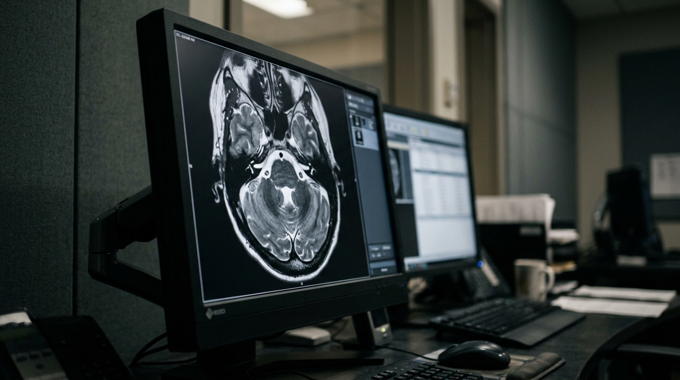
A systematic review and meta-analysis evaluating magnetic resonance imaging artificial intelligence models has found that these quantitative tools demonstrate moderate discriminative accuracy when identifying pain-related nerve phenotypes in patients with non-secondary trigeminal neuralgia. The synthesis, published by researchers from The Fifth Affiliated Hospital of Zhengzhou University in Frontiers in Radiology, evaluated pooled diagnostic metrics across eight eligible investigations comprising 628 nerve-level observations. Applying a bivariate random-effects statistical model, the research team established that the evaluated machine-learning architectures achieved a pooled sensitivity of 0.83 with a 95 percent confidence interval extending from 0.78 to 0.87, accompanied by a pooled specificity of 0.81 with a 95 percent confidence interval spanning 0.74 to 0.86. The corresponding summary receiver operating characteristic curve yielded an area under the curve of 0.86 with a 95 percent confidence interval between 0.82 and 0.88, demonstrating stable discriminatory capacity across varied imaging environments while functioning primarily as potential adjunctive diagnostic instruments.
Trigeminal neuralgia represents a debilitating orofacial neuropathic disorder marked by recurrent, brief, electric shock-like or lancinating pain paroxysms that affect one or more divisions of the trigeminal nerve. These painful episodes are frequently precipitated by innocuous everyday stimuli, including light facial touch, eating, or speaking, and often impose severe detriments on patient sleep quality and overall daily living. Within clinical diagnostic frameworks, trigeminal neuralgia is systematically divided into classical, secondary, and idiopathic etiologies. The investigators adopted the operational term non-secondary trigeminal neuralgia to specifically encompass classical and idiopathic presentations once secondary structural causes, such as compressive intracranial tumors or multiple sclerosis plaques, have been formally ruled out. Precise discrimination between these neuropathic classifications remains pivotal for guiding clinical interventions, as anatomical configurations and neurovascular relationships directly shape clinical judgment regarding suitability for neurosurgical procedures such as microvascular decompression.
Standard diagnostic workups depend heavily on conventional visual assessment of magnetic resonance imaging scans to identify physical conflicts between cranial blood vessels and the trigeminal root. Nevertheless, visual assessment exhibits substantial clinical limitations because simple physical contact between a vessel and the nerve is not uniquely symptomatic. Routine neurovascular contact is frequently detected in entirely asymptomatic individuals as well as along the asymptomatic contralateral trigeminal nerves of individuals suffering from unilateral trigeminal neuralgia. Standard clinical criteria established by the International Classification of Headache Disorders, Third Edition, and the International Classification of Orofacial Pain dictate that classical trigeminal neuralgia requires neurovascular compression accompanied by definite morphological alterations, including nerve displacement or cross-sectional atrophy, rather than incidental contact alone. Even when utilizing high-resolution magnetic resonance sequences, standard radiologic visual inspections remain constrained by human observer subjectivity, subtle venous compressions, diminutive offending vessels, and intricate anatomical variations.
The biological rationale for adopting objective quantitative neuroimaging methods extends beyond the macroscopic visualization of macroscopic vascular compression. In classical trigeminal neuralgia, focal demyelination, local axonal impairment, and hyperexcitability near the root entry zone can promote ectopic impulse generation and ephaptic cross-talk between sensory fibers. Furthermore, while idiopathic trigeminal neuralgia lacks the conspicuous neurovascular morphological distortions characteristic of classical disease, specialized structural and diffusion magnetic resonance examinations have repeatedly uncovered subtle quantitative alterations that escape standard visual detection. Radiomic feature analysis and modern machine-learning models address this gap by extracting high-dimensional mathematical data from medical images, measuring complex shape geometries, gray-level intensity variations, texture heterogeneity, and spatial filtering profiles. When integrated into classification pipelines, these algorithmic frameworks synthesize objective numerical representations that can help classify affected nerve sides without replacing direct clinical histories or standard etiological categorizations.
To rigorously aggregate existing evidence, the review team executed a systematic literature search adhering to the Preferred Reporting Items for Systematic Reviews and Meta-Analyses 2020 statement and its diagnostic test accuracy extension, registering their protocol prospectively in the PROSPERO registry under identification number CRD420261394456. Candidate studies were screened against strict inclusion rules requiring enrolled cohorts to possess verified non-secondary trigeminal neuralgia diagnosed via international criteria, index models designed to classify affected nerves against valid comparators, and sufficient documented data to populate complete two-by-two diagnostic contingency tables reflecting validation or cross-validation test performance.
The comprehensive bibliographic queries initially recovered 164 total citations, which yielded 103 unique publications after the removal of 61 duplicate records. Subsequent title and abstract screening eliminated 65 unsuitable references, leaving 38 full-text reports for exhaustive methodological appraisal. Ultimately, eight studies met every predefined eligibility standard and were enrolled in the quantitative meta-analysis, contributing a cumulative total of 628 evaluated nerve units. Two of the eight studies supplied direct extractable contingency tables, two allowed exact derivation from reported cohort statistics, and four required approximate numerical reconstruction from validation averages.
Methodological characteristics across the eight included studies revealed varied engineering workflows, classification algorithms, and magnetic resonance protocols. Six investigations utilized 3.0-Tesla scanners exclusively, one utilized a 1.5-Tesla system, and one incorporated both field strengths across patient groups. Manual delineation of regions of interest was performed in six studies, whereas two investigations implemented automated segmentation procedures. With respect to input protocols, three investigations relied on single magnetic resonance sequences, while five extracted predictive features across multiple sequences, such as three-dimensional volumetric interpolated breath-hold examination, short tau inversion recovery, and fast low-angle shot imaging. In terms of algorithmic architectures, models ranged from logistic regression classifiers and support vector machines to shallow artificial neural networks, Gaussian processes, and subspace discriminant ensembles, with individual cohort evaluations reporting sensitivities between 0.73 and 1.00 and specificities spanning from 0.67 to 0.97.
Methodological quality and risk of bias were evaluated utilizing the Quality Assessment of Diagnostic Accuracy Studies 2 tool alongside the specialized Prediction Model Risk of Bias Assessment Tool for regression and artificial intelligence models. Under the diagnostic accuracy appraisal, one study displayed high risk of bias regarding index test execution because interpreting radiologists possessed knowledge of clinical laterality during nerve segmentation. Low risk regarding classification thresholds was confirmed in four studies that locked predictive decision rules exclusively within training partitions prior to testing. When examined under artificial intelligence quality criteria, three studies exhibited high risk of bias within the analytical domain during model construction, and three showed high risk within validation evaluations. The identified limitations stemmed primarily from small evaluation cohorts, inadequate external verification, and potential vulnerabilities surrounding data leakage during feature optimization, prompting recommendations for cautious interpretation of model performance.
In the primary statistical synthesis, the bivariate mixed-effects model demonstrated mild statistical heterogeneity across the eight evaluated studies. Calculated inconsistency values were 20.77 percent with a 95 percent confidence interval of 0.00 to 81.23 percent for pooled sensitivity, and 27.65 percent with a 95 percent confidence interval of 0.00 to 85.50 percent for pooled specificity. An exploratory Spearman correlation between the logit of sensitivity and the logit of false-positive rates yielded a correlation coefficient of 0.119 with a p-value of 0.775, confirming no detectable threshold effect. In leave-one-out sensitivity testing, excluding an influential study by Cüce and colleagues shifted the pooled sensitivity to 0.82, adjusted specificity to 0.78, and established a revised summary area under the curve of 0.87, while reducing the heterogeneity metric for specificity to zero percent.
The meta-analysis authors conducted exploratory subgroup and sensitivity evaluations to test whether specific modeling choices altered discriminatory accuracy. When stratified by analytical feature strategy, four investigations employing radiomics features alone demonstrated a pooled sensitivity of 0.84, a pooled specificity of 0.84, and a summary area under the curve of 0.89. In comparison, four studies that combined radiomic signatures with macro-anatomical or morphological variables achieved a pooled sensitivity of 0.86, a pooled specificity of 0.78, and an area under the curve of 0.88. An exploratory analysis restricted to the five studies utilizing healthy-control comparison groups demonstrated summary sensitivity of 0.82, summary specificity of 0.85, and an area under the curve of 0.90. Similarly, restricting analysis to the four studies possessing directly reported or uniquely derivable contingency tables yielded a summary sensitivity of 0.82 and a specificity of 0.81.
Multiple critical methodological caveats constrain the broader clinical translation of the current evidence base. Foremost, the aggregate evidence relies upon only eight studies that were predominantly single-center, retrospective investigations conducted in relatively small samples without independent external multicenter testing. Additionally, combining within-patient contralateral nerve designs with separate healthy-control comparisons merged two biologically disparate tasks, since contralateral nerves introduce within-patient anatomical correlation that published aggregate data could not account for via cluster-adjusted modeling. Funnel-plot asymmetry testing using Deeks method yielded a p-value of 0.10, indicating that while formal asymmetry was not statistically confirmed, small-study effects and publication bias cannot be excluded given the modest sample size. Furthermore, literature retrieval was confined to medical databases without searching dedicated computing platforms such as IEEE Xplore, potentially missing relevant engineering contributions.
The study authors concluded that while magnetic resonance imaging radiomics and artificial intelligence algorithms demonstrate moderate discriminative capacity for identifying affected nerve phenotypes in non-secondary trigeminal neuralgia, current evidence remains preliminary. The observed metrics indicate that algorithmic predictions should be interpreted purely as quantitative image characterizations rather than definitive evidence of causal pathophysiology or clinical utility. Machine-learning models are not designed to supplant detailed patient interviews, clinical pain histories, or expert neurosurgical interpretation of neurovascular relationships. Future investigations must prioritize prospective multicenter cohorts featuring transparent algorithmic architectures, automated segmentation, compliance with Image Biomarker Standardisation Initiative benchmarks, independent external validation sets, and rigorous adjustment for paired nerve-level observations before algorithmic diagnostic platforms can transition into standard clinical workflows.
Subject of Research: Diagnostic performance of MRI-based artificial intelligence models in non-secondary trigeminal neuralgia
Article Title: Magnetic resonance imaging-based artificial intelligence in identifying imaging phenotypes of non-secondary trigeminal neuralgia: a systematic review and meta-analysis
Article References: Huo, X., Li, J., Wang, B., Zhao, T., Wu, H., Cheng, S., Wu, M., Gao, H., Liu, R., Wang, J., & Shou, J. (2026). Magnetic resonance imaging-based artificial intelligence in identifying imaging phenotypes of non-secondary trigeminal neuralgia: a systematic review and meta-analysis. Frontiers in Radiology, 6, Article 1980185. https://doi.org/10.3389/fradi.2026.1980185
Image Credits: AI Generated
DOI: 10.3389/fradi.2026.1980185
Keywords: trigeminal neuralgia, artificial intelligence, magnetic resonance imaging, radiomics, systematic review, meta-analysis, diagnostic accuracy, neuroimaging, Magnetic, resonance, imaging-based, artificial
News Source: Blake Davidson. (November 6, 2026). Artificial Intelligence Models Show Moderate Accuracy in Detecting Trigeminal Neuralgia Phenotypes. Scienmag.